Contents
Cell Structure
Cell Structure
There are two main types of cells: eukaryotic and prokaryotic cells. Eukaryotic cells are larger than prokaryotic cells and their DNA is contained in a nucleus.
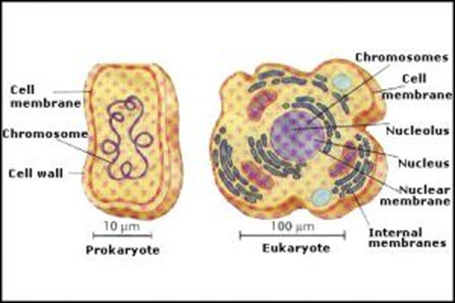
Most eukaryotic cells are complex multicellular organisms containing a range of specialized cells to perform a variety of functions. Organisms are made up of organ systems, which contain a range of organs working together to perform a function. Organs are made up of a range of tissues working together to perform a function, and specialized cells of a similar structure and function are organized into tissues.
Eukaryotic Cells
Eukaryotic cells contain a range of organelles.
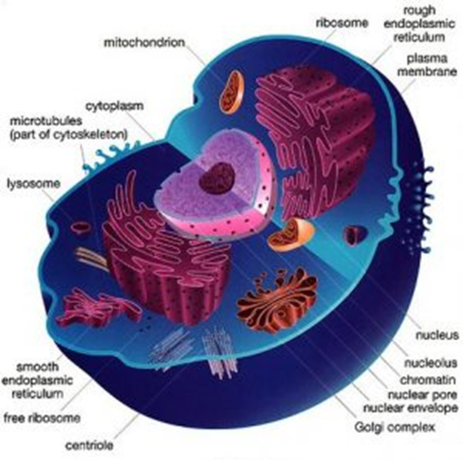
| Organelle | Structure | Function |
| Cell-surface membrane | Phospholipid bilayer with proteins and cholesterol embedded within it. Glycolipids and glycoproteins in the surface. | The fluid-mosaic model of the membrane refers to the fluidity and range of molecules in the membrane. Cholesterol provides strength and reduces fluidity, proteins are for transport, and the glycoproteins and glycolipids are for cell recognition and act as receptors. |
| Nucleus | Surrounded by a double membrane nuclear envelope with nuclear pores. Containing chromosomes, consisting of protein-bound, linear DNA, and one or more nucleolus | Nucleolus is the site of rRNA production and makes ribosomes. DNA replication and transcription occur in the nucleus. |
| Mitochondria | Double membrane organelle. The inner membrane is folded to form cristae. Contains a fluid center called the matrix | Site of aerobic respiration and ATP production. |
| Chloroplasts | Surrounded by a double membrane. Contains thylakoids, which are folded membranes containing chlorophyll pigments. Contains a fluid center, the stroma. | The site of photosynthesis. The stroma contains enzymes for the light-independent stage of photosynthesis. |
| Golgi apparatus and Golgi vesicles | Stacks of membranes creating flattened sacs called cisternae, surrounded by small round hollow vesicles. | Proteins and lipids are modified here. Carbohydrates can be added to proteins to form glycoproteins. Finished products are transported in the Golgi vesicles. |
| Lysosomes | Formed when the Golgi apparatus contains hydrolytic enzymes. | A type of Golgi vesicle that releases lysozymes. |
| Ribosomes | Small granules in cells made of protein and rRNA. Made up of a small and large subunit. 80s in size in eukaryotes. | The site of translation in protein synthesis. |
| Rough endoplasmic reticulum (RER) and smooth endoplasmic reticulum (SER) | Sheets of membranes linked to the nucleus. The membranes form a network of cisternae - a network of tubules and flattened sacs. | The RER has ribosomes on the outer surface. RER - site of protein synthesis and glycoprotein synthesis. The proteins can then be transported through the RER. SER – create, store, and transport lipids and carbohydrates. |
| Cell wall | Found in plants, algae, and fungi. Consists of polysaccharides, cellulose in plants and chitin in fungi. There is a thin boundary layer between adjacent cells called the middle lamella. | Provides structural strength to cells and prevents cells from bursting when water enters by osmosis. |
| Cell vacuole | Found in plants. A single membrane sac filled with fluid containing salts, sugars, and amino acids. The membrane around a cell vacuole is called the tonoplast. | To provide support to a cell, store amino acids and sugars, and can contain pigments to attract pollinators. |
| Cytoskeleton | Found within the cytoplasm all over a cell. | Provides mechanical strength to cells, helps with transport within cells, and allows cell movement. Many organelles are bound to the cytoskeleton. |
Prokaryotic Cells
Bacterial cells are the key example of prokaryotic cells. In addition to them being smaller and not having a nucleus, there are other differences compared to a eukaryotic cell shown in the diagram below.
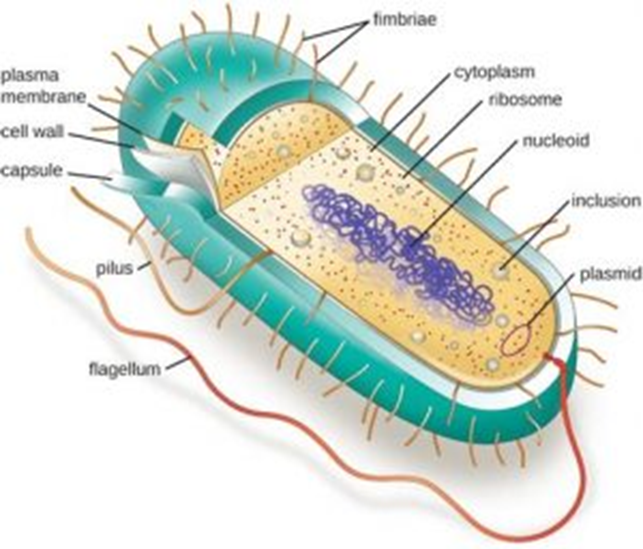
The cytoplasm does not contain any membrane-bound organelles, and the ribosomes are smaller, 70s in size. Bacteria still contain DNA, but it is not stored in a nucleus. The DNA is found as a single, circular molecule in the cytoplasm and it is not associated with histone proteins.
Prokaryotic cells do have cell walls, but they do not contain cellulose or chitin. Instead, they are made of a glycoprotein called murein.
Some bacteria also contain three additional features. They can contain plasmids, which are rings of DNA containing genes linked to survival such as antibiotic resistance. Surrounding the cell wall, some bacteria have a capsule which is to provide protection from other cells and to help bacteria stick together. Finally, they can have flagella which are used for locomotion.
Methods of Studying Cells
Knowing the internal structure of cells has been possible due to the use of microscopes.
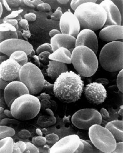
Some cell components and cells are difficult to see unless they are stained a more obvious color. Differential staining is a technique that can be used, which involves many chemical stains being used to stain different structures different colors. A common application of this in medicine is to detect abnormalities in the proportion of the different white blood cells in a patient’s blood sample.
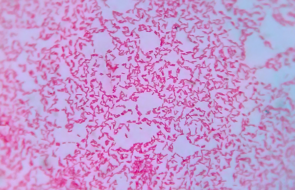
Gram staining, to visualize different bacteria, is another common use of differential staining. Two different stains are used, crystal violet and Fuchsin/Safranin, to enable Gram-positive and Gram-negative bacteria to be differentiated under the microscope. Gram-positive and Gram-negative have different thickness cell walls. Gram-positive bacteria can take up the crystal violet stain and appear purple under the microscope. This is due to the thick peptidoglycan cell wall layer absorbing the dye.
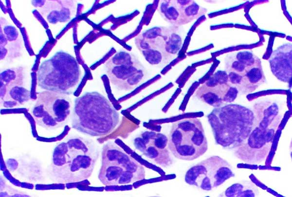
Stained Gram-positive bacteria (long dark rods) and WBC with the lobed nucleus stained.
On the contrary, Gram-negative bacteria cannot absorb crystal violet stain as their peptidoglycan cell wall is thin. Being able to distinguish between the two types of bacteria helps medics to prescribe an appropriate antibiotic to patients with bacterial infections.
Gram-negative bacteria stained red.
Microscopes
There are four key types of microscopes: optical microscopes, transmission electron microscopes, scanning electron microscopes, and laser scanning confocal microscopes.
The magnification of a microscope refers to how many times larger the image is compared to the object.
The resolution of a microscope is the minimum distance between two objects in which they can still be viewed as separate. The resolution in an optical microscope is determined by the wavelength of light, and the wavelength of the beam of electrons determines the resolution in an electron microscope.
Optical (Light) Microscopes
Light microscopes have a poor resolution due to the long wavelength of light. Small organelles in a cell are not visible using an optical microscope, but living samples can be examined and a color image is obtained.
Structures viewed under an optical microscope can be measured using the formula:
magnification = size of image / size of real object
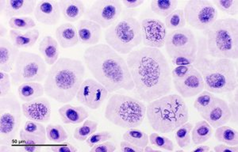
Cells undergoing mitosis shown with an optical microscope.
Observations of structures from under a microscope can be recorded in the form of scientific drawings.
There are a few rules to scientific drawings, which makes them very different from artistic drawings:
- Draw in pencil
- Title the diagram to indicate what the specimen is
- State the magnification that you are drawing it from
- Annotate cell components, cells, and sections of tissue visible
- Do not sketch. Only use solid lines that do not overlap.
- Do not color in or shade.
The aim is to show size, location, and proportion.
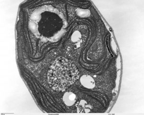
Electron Microscopes (EM)
A beam of electrons has a very short wavelength and therefore EMs have a high resolution and small organelles can be visualized. An image is created using an electromagnet to focus the beam of negatively charged electrons.
Electrons are absorbed by air, which is why samples for an EM must be in a vacuum. For this reason, only non-living specimens can be examined. The image is also black and white as the samples must be stained.
Transmission Electron Microscopes (TEM)
Extremely thin specimens are stained and placed in a vacuum. An electron gun produces a beam of electrons that pass through the specimen. Some parts of the specimen absorb the electrons and this makes them appear dark. The image produced is 2D and shows detailed images of the internal structure of cells.
Scanning Electron Microscope (SEM)
The specimens do not need to be thin, as the electrons are not transmitting through. Instead, the electrons are beamed onto the surface and the electrons are scattered in different ways depending on the contours. This produces a 3D image.
Confocal laser scanning fluorescence micrograph of part of a plant stamen.
- Which cell components are only sometimes found in prokaryotic cells?
- Your answer should include: Flagellum / Plasmid / Pili / Capsule
- What are the four types of microscopes?
- Your answer should include: Light / Optical / Electron / Transmission / Laser / Scanning / Confocal
- What is the formula for magnification?
- Your answer should include: Magnification / Image / Actual / Size
- Do electron or light microscopes have a higher resolution?
- Electron
- Which organelle is the site of protein synthesis?
- Your answer should include: Ribosome / RER
- Which organelle is where proteins are modified and secreted from?
- Your answer should include: Golgi / Apparatus