Contents
Cell Division, Cell Diversity, and Cellular Organization
Cell Division
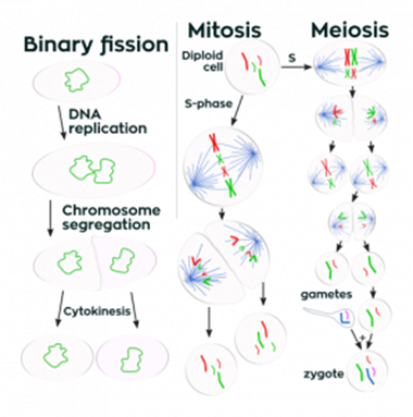
Eukaryotic cells enter the cell cycle and divide by mitosis or meiosis. In comparison, prokaryotic cells replicate by binary fission, and viruses do not undergo cell division as they are non-living. Viruses replicate inside the host cells they invade by injecting their nucleic acid into the cell to replicate the virus particles.
Stem Cells
Multicellular organisms have a diverse range of specialized cells that all originate as undifferentiated stem cells. Stem cells are undifferentiated cells that can continually divide and become specialized.
Differentiation is the process by which stem cells become specialized. The key specialized cells you need to be able to describe the structure of and link to their function are:
- Erythrocytes - biconcave shape to increase surface area for diffusion and increase flexibility to fit through narrow capillaries. No nucleus so there is more space to hold hemoglobin to increase oxygen transport.
- Neutrophils - Flexible cell to enable them to surround pathogens and engulf them. They contain lysosomes filled with the hydrolytic enzyme, lysozyme.
- Squamous epithelial cells - Usually only a single layer of flat cells in contact with the basement membrane of the epithelium. This provides a short diffusion distance.
- Ciliated epithelial cells - These cells have hair-like projections that sway to move substances, such as mucus in the lungs or an egg in the oviduct.
- Sperm cells - Flagellum containing many mitochondria to release energy for locomotion and contains digestive enzymes in the tip of the head to digest the wall of the egg cell.
- Palisade cells - Contain many chloroplasts to maximize light energy absorption for photosynthesis.
- Root hair cells - Increase surface area for osmosis of water and active transport of minerals due to the projection of the cell. Has a thin cell wall to reduce the diffusion distance.
- Guard cells - Flexible walls, more so on one side, which results in the cells bending when turgid to open stomata and close when flaccid, helping to control water loss.
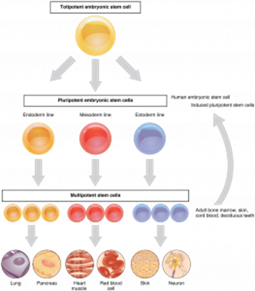
There are different types of stem cells that have different differentiation abilities. These are totipotent, pluripotent, multipotent, and unipotent stem cells.
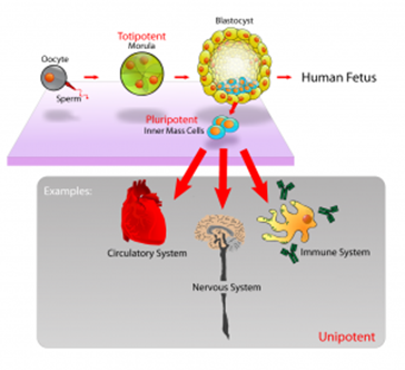
Totipotent cells can divide and produce any type of body cell. During development, totipotent cells translate only part of their DNA, resulting in cell specialization. Totipotent cells occur only for a limited time in early mammalian embryos.
Pluripotent cells are found in embryos and can become almost any type of cell. For this reason, they are used in research with the prospect of using them to treat human disorders. These stem cells could be used to regrow damaged cells in humans, such as replacing burnt skin cells or beta cells for type I diabetics or neurons in Parkinson’s disease sufferers. There are issues with this as sometimes the treatment doesn’t work, or the stem cells continually divide to create tumors. Additionally, ethically there is debate on whether it is right to make a therapeutic clone of yourself to make an embryo to get the stem cells to cure a disease and then destroy the embryo. Is it safe or even sensible to clone humans and destroy embryos?
Multipotent and unipotent cells are found in mature mammals and can divide to form a limited number of different cell types. Multipotent cells, such as in bone marrow, can differentiate into a limited number of cells such as erythrocytes and neutrophils, whereas unipotent cells can only differentiate into one type of cell.

The stem cells in adult plants are found at meristems. This is where the production of tissues, such as xylem and phloem sieve tubes, is formed.
Sources of Stem Cells in Mammals:
Embryos, up to 16 days after fertilization, contain stem cells that are pluripotent and can differentiate into any type of cells.
Umbilical cord blood contains stem cells that are multipotent, like adult stem cells.
The placenta has stem cells that can develop into a limited number of specialized cells.
Adult stem cells, such as in the bone marrow, can produce different cells to repair those within a particular tissue or organ.
There are pros and cons of using stem cells to treat human disorders. The benefits would include research or even treatment of damaged tissues for neurological conditions like Alzheimer’s and Parkinson’s, and research into developmental biology. Induced pluripotent stem cells (iPS cells) can be produced from adult somatic cells using appropriate protein transcription factors to overcome some of the ethical issues with using embryonic stem cells.
iPS cells are created from adult unipotent cells. These cells, which can be from almost any body cell, are altered in the lab to return them to a state of pluripotency. To do this, the genes that were switched off to make the cell specialized must be switched back on. This is done using transcription factors. They are very similar to embryonic pluripotent stem cells but do not cause the destruction of an embryo, and the adult can give permission. Also, the iPS have shown a self-renewal property, in that they can divide indefinitely to give limitless supplies. For these reasons, they could be used in medical treatment instead of embryonic stem cells.
- List the three main stages of the cell cycle:
- Your answer should include: Interphase / Nuclear / Division / Cytokinesis
- When does DNA replication occur?
- Your answer should include: S Phase / S-phase / Interphase
- Which stem cells can differentiate into any type of cell?
- Totipotent
- Which is the longest stage of the cell cycle?
- Interphase
The Cell Cycle
The cell cycle comprises three key stages: interphase (G1, S, G2), nuclear division (mitosis or meiosis), and cytokinesis.
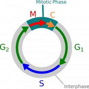
Interphase is the longest stage in the cell cycle. Interphase is when the organelles double, the cell grows, and then DNA replicates.
Nuclear division can be either mitosis, creating two identical diploid cells, or meiosis, creating four genetically different haploid cells. Mitosis creates cells with identical DNA for growth and repair, whereas meiosis creates gametes.
Cytokinesis is the final stage. It is the division of the cytoplasm to create the new cells.
Mitosis
Mitosis has four key stages: prophase, metaphase, anaphase, and telophase.
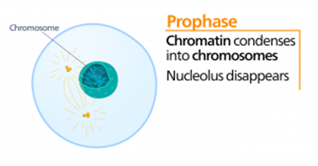
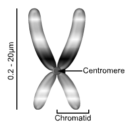
Prophase
In this stage, the chromosomes condense and become visible. In animal cells, the centrioles separate and move to opposite poles of the cell. The centrioles are responsible for creating spindle fibers which are released from both poles to create a spindle apparatus – these will attach to the centromere and chromatids on the chromosome in later stages. Plants have a spindle apparatus but lack the centrioles.
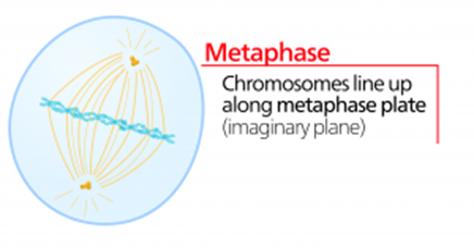
Metaphase
The chromosomes align along the equator of the cell. The spindle fibers released from the poles now attach to the centromere and chromatid.
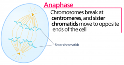
Anaphase
The spindle fibers start to retract and pull the centromere and chromatids they are bound to towards the opposite poles. This causes the centromere to divide in two, and the individual chromatids are pulled to each opposite pole. These separated chromatids are now referred to as chromosomes.
This stage requires energy in the form of ATP, which is provided by respiration in the mitochondria.
Telophase
The chromosomes are now at each pole of the cell and become longer and thinner again. The spindle fibers disintegrate, and the nucleus starts to reform.
The final stage in the cell cycle is when the cytoplasm splits in two to create the two new genetically identical cells.
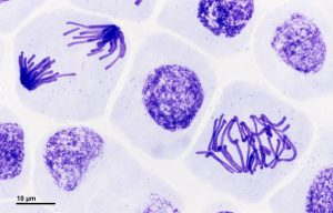
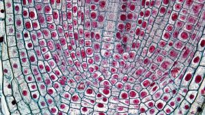
The stages of mitosis are visible under a light microscope in onion and garlic root tips. The tips of the roots are constantly dividing for growth, and therefore many of the cells are replicating, and the different stages of the cell cycle and mitosis are visible. A small slice of the root tip is placed on a microscope slide and broken down with a needle. A stain is added to make the chromosomes visible, and the cover slip is pushed down. This is to squash the tip to achieve a single layer of cells so it is possible to focus on the cells. The mitotic index can be calculated by counting how many cells are visible in the field of view and the number of cells visible that are in a stage of mitosis.
Then the following formula can be used:
Mitotic index = (the number of cells in mitosis / the total number of cells) x 100
Genetic Variation
This is introduced in a variety of ways, such as during meiosis cell division, mutations, and the random fertilization of gametes.
Meiosis
This is the type of cell division that creates genetically different gametes. Unlike in mitosis, there are two nuclear divisions in this process, which results in four haploid daughter cells.
Haploid (n) = one copy of each chromosome
Diploid (2n) = two copies of each chromosome
The genetic differences are introduced by two key processes in meiosis:
- Independent segregation of homologous chromosomes
- Crossing over between homologous chromosomes
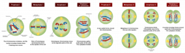
The above diagram outlines the stages in meiosis. Before cell division, interphase occurs in which the DNA and organelles double.
As meiosis involves two nuclear divisions, the descriptions of the processes can be stated as being in meiosis I or meiosis II, referring to the stage of nuclear division. Both stages include prophase, metaphase, anaphase, telophase, and cytokinesis.
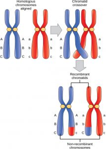
Crossing Over
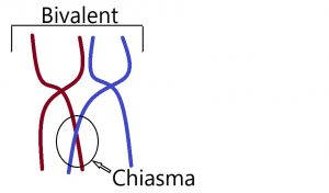
During prophase I (one), the chromosomes condense and become thicker. The homologous chromosomes pair to form bivalents. Crossing over of genetic material can occur between the chromatids of bivalents. Where the chromatids cross over is called the chiasma - there can be more than one chiasma at a time. Breaks can occur in the genetic material, and parts of the chromatids are exchanged between the homologous pairs. This results in new combinations of alleles in the resulting gamete.
Independent Segregation
During metaphase I, in meiosis I, the homologous pairs of chromosomes line up opposite each other on either side of the equator. As there are 23 different homologous pairs, there are 8,388,608 different ways the pairs could assort themselves (2 to the power of 23). It is random each time which side of the equator the paternal and maternal chromosomes of each homologous pair align at the equator, and as a result, each gamete receives different combinations of the maternal and paternal chromosomes.