Contents
Biological Molecules
Carbohydrates
Carbohydrates are essential biological molecules that store energy and provide structural support to plant cells. Carbohydrates can be classified into three groups based on the number of units they consist of, as shown in the flow diagram below.
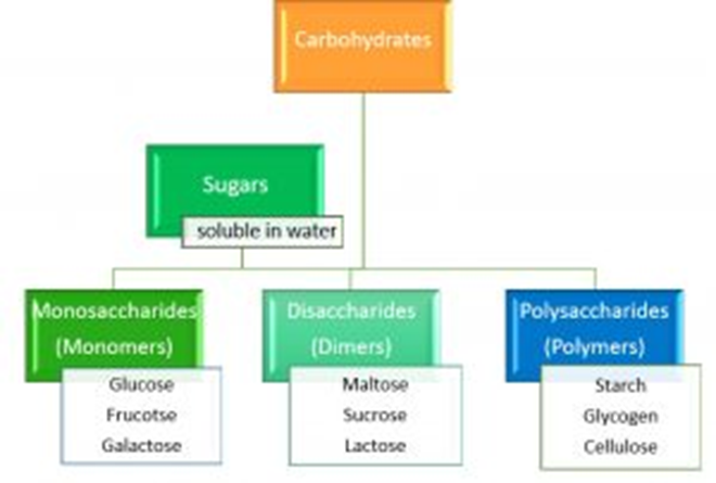
Larger carbohydrates, such as sucrose and starch, are composed of monosaccharides. The individual units of carbohydrates are known as monosaccharides, with glucose, galactose, and fructose being common examples. Monosaccharides are all water-soluble sugars and serve either as an energy source or as building blocks for other molecules.
All carbohydrates consist of three elements: carbon, hydrogen, and oxygen (CHO). The general formula for a monosaccharide is CnH2nOn, where n is the number of carbon atoms it contains.
Glucose
Glucose, C6H12O6, is a crucial monosaccharide that can provide energy, polymerize to form structural molecules (cellulose), or serve as an energy storage molecule (glycogen and starch). It contains six carbon atoms, highlighted in red in diagram (a). You may be required to draw glucose, but fortunately, only in as much detail as shown in diagram (b).
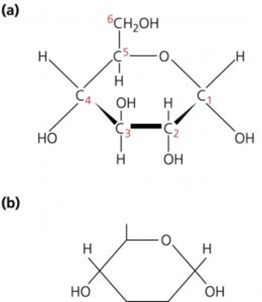
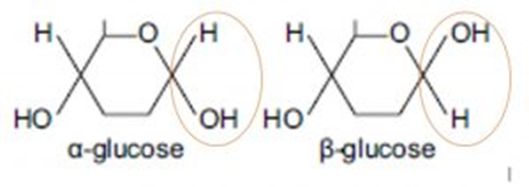
Glucose has two structural isomers (isomers are compounds with the same formula but different atom arrangements). The diagrams above depict the isomer α glucose. β glucose is the second isomer. The only difference in structural arrangement between these isomers is found on carbon atom 1 in the diagram below, where the positions of hydrogen (H) and the hydroxyl group (OH) have swapped. This small change significantly impacts the bonding and final structure of the polymers they form.
- α glucose and β glucose only differ structurally on one of their carbon atoms. Which carbon atom is it where H and OH swap positions?
- Your answer should include: 1 / One
- Glucose is a vital monosaccharide with multiple functions. What are its functions?
- Your answer should include: Energy / Storage / Structural / Support
- Name three common monosaccharides:
- Your answer should include: Glucose / Galactose / Fructose
- Which types of carbohydrates are categorized as sugars?
- Your answer should include: Monosaccharides / Disaccharides
Monomers and Polymers
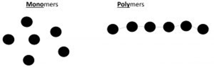
Monomers (mono meaning one, think monobrow!)
Monomers are small, single units that act as building blocks to create larger molecules.
Polymers (poly meaning more than two)
Polymers are composed of many monomers, often thousands, chemically bonded together.
For monomers to bond together, a chemical reaction occurs, which is a condensation reaction. Condensation reactions involve the removal of water. This removal of water from monomers enables a chemical bond to form between them.
A hydrolysis reaction is the reverse of this process. Hydrolysis (hydro meaning water, lysis meaning to split) involves the addition of a water molecule between two bonded monomers (within a dimer or polymer) to break the chemical bond.

Disaccharides
- Formed from two monosaccharides
- Joined by a glycosidic bond
- Formed by a condensation reaction
There are three key disaccharides that you need to remember, and these are made from the three key monosaccharides you learned:
- Glucose + Glucose –> Maltose
- Glucose + Galactose –> Lactose
- Glucose + Fructose –> Sucrose
The general formula for a disaccharide is:
(CnH2nOn) 2 – H20
Where n = the number of carbon atoms the monosaccharide contains. This formula is simply twice the monosaccharide formula minus water.
Condensation Reaction
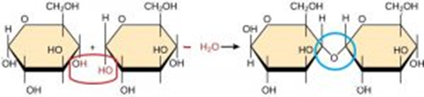
The diagram below demonstrates how a condensation reaction creates a disaccharide. A water molecule is being removed (highlighted in red) from the hydroxyl group (OH) on carbon 1 and carbon 4 on the two monosaccharides. The bond that forms is known as a glycosidic bond (highlighted in blue). This diagram shows a 1,4-glycosidic bond because it is located between carbon 1 and carbon 4.
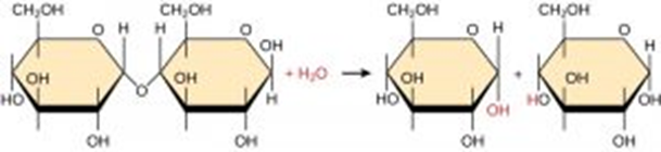
Disaccharides can be broken down back into monosaccharides via a hydrolysis reaction. Hydrolysis is when the water that was removed is added back again to break the glycosidic bond, as can be seen in the diagram below.
Reducing and Non-Reducing Sugars Test
Both monosaccharides and disaccharides are described as sugars because they are sweet and soluble. An experiment can be conducted to test for the presence of these sugars. All of these sugars, except sucrose, are reducing sugars. Sucrose is a non-reducing sugar.
Reducing Sugar Test
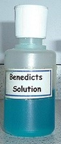
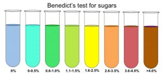
To test for the presence of these sugars, Benedict’s reagent is added. This reagent is a bright blue liquid due to its copper sulfate content. The term "reducing sugar" is given to sugars that can reduce Cu2+ ions in Benedict’s reagent to Cu+ ions, forming copper (I) oxide, which results in a brick red precipitate.
The chemical procedure for this is:
- Add Benedict’s reagent to the sample you are testing
- Heat
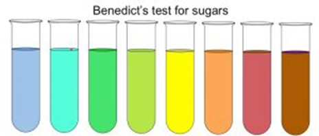
- If a color change from blue to yellow/green/red is observed, then this confirms the presence of a reducing sugar. The more red/brown the precipitate, the more sugar it contains.
Non-Reducing Sugar Test
Sucrose is called a non-reducing sugar because it cannot reduce Cu2+. This is because the chemical group needed for this reduction reaction is involved in the glycosidic bonds between the monosaccharides. To prove that sucrose is still a sugar but is just unable to reduce Cu2+ (a non-reducing sugar), the glycosidic bond must be hydrolyzed to expose the reducing group.
If a substance remains blue after the reducing sugars test, then the procedure to test to see if it is a non-reducing sugar is as follows:
- Mix sucrose with HCl and boil. This is acid hydrolysis and it breaks the glycosidic bond so that sucrose is hydrolyzed back into glucose and fructose. Hint: To get the mark, you must state "BOIL," as below 100°C there is not enough energy to break the glycosidic bond.
- Cool the solution and then add sodium hydroxide to make the solution alkaline. Benedict’s reagent only works in alkaline solutions, which is why this stage is essential. You must cool the solution first to prevent excessive, dangerous fizzing.
- Add a few drops of Benedict’s reagent and heat.
- If a color change from blue to yellow/green/red is observed, then this is confirmation that a non-reducing sugar is present.
Polysaccharides
Polysaccharides are polymers made up of many monosaccharides. A polysaccharide is created in the same way as a disaccharide, via condensation reactions.
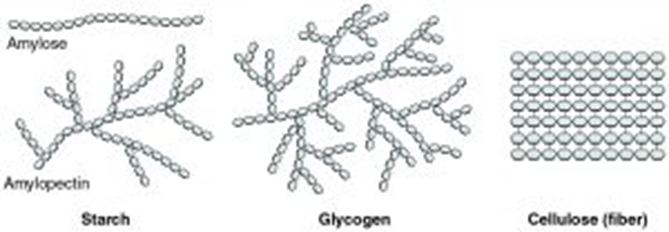
There are three key polysaccharides that you need to learn the structure and function of: starch, glycogen, and cellulose. Starch and glycogen are both energy stores, whereas cellulose provides structural support.
Starch
Starch is found in plants, not in animal cells, and it is the major carbohydrate store. Starch is made from the excess glucose created during photosynthesis. Glucose is used in respiration, but if more glucose is created in photosynthesis than is currently needed, it is converted into the polymer starch for storage.
The presence of starch can be confirmed by using iodine. Iodine is orange/brown in color when no starch is present, but it turns blue/black if starch is present.

Structure of Starch
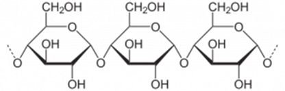
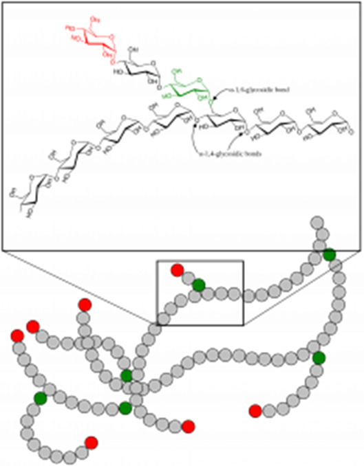
Starch is a polymer made up of α-glucose. These α-glucose monomers are joined together via condensation reactions and are held in place by 1-4 and 1-6 glycosidic bonds (remember, the numbers refer to which carbon atoms the bond forms between).
Amylose is the name of the structure in starch in which the glucose monomers are all joined together by 1,4-glycosidic bonds. This results in a spiral-shaped polymer, as seen in the diagram below.
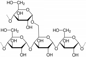
Amylopectin is the name given to the other structure in starch in which the glucose monomers are joined by a combination of 1,4 and 1,6 glycosidic bonds. The 1,6-glycosidic bonds result in branches, as shown in the diagram below.
Amylose
Amylopectin
Properties of Starch
Starch is insoluble due to its large molecule size. This is advantageous for a storage molecule as it can be stored within cells without dissolving, thus not affecting the water potential of a cell or osmosis.
The spiral shape of amylose allows for efficient compaction, enabling a large portion of the molecule to fit into small spaces for storage.
The branching strands in amylopectin provide a larger surface area for enzymes to attach to. This makes starch readily hydrolyzed back into glucose when plant cells are running low on glucose.
Glycogen
Glycogen is the major carbohydrate storage molecule found in animal cells, primarily in liver and muscle cells. Glycogen is created from excess glucose absorbed into the bloodstream after eating.
Glucose is used in respiration, and any excess glucose is converted into the polymer glycogen for storage. Liver cells, responsible for detoxification, and muscle cells, responsible for movement, store glycogen to ensure they have a glucose reserve for energy.
Structure of Glycogen
Glycogen is a polymer made up of α-glucose and shares a similar structure with amylopectin in starch. α-Glucose monomers are joined through condensation reactions and held together by 1,4 and 1,6-glycosidic bonds. The key distinction is that glycogen has more 1,6-glycosidic bonds, resulting in a highly branched structure.
Properties of Glycogen
Glycogen's large molecule size makes it insoluble, allowing for storage within cells without affecting the water potential or osmosis.
Due to its highly branched structure, glycogen provides a larger surface area for enzymes to attach to. Consequently, it is rapidly hydrolyzed back into glucose when cells require a quick energy source. This is crucial for animals with higher metabolic rates, such as fleeing from predators.
Water
The Structure of Water
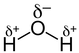
Water is a dipolar molecule, with oxygen being slightly negative and hydrogen atoms being slightly positive. The delta (δ) symbol indicates this slight positive/negative charge on the diagram below.
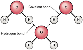
Hydrogen bonds form between different water molecules, connecting the oxygen and hydrogen atoms.
The formation of these hydrogen bonds and water's dipolarity result in five key properties of water.
Five Key Properties of Water
- It is a metabolite, playing a role in condensation and hydrolysis reactions.
- It is an important solvent in various reactions.
- Water has a high heat capacity, providing temperature buffering.
- It has a significant latent heat of vaporization, creating a cooling effect when water evaporates.
- Strong cohesion exists between water molecules, supporting water columns and providing surface tension.
Metabolite
Water is involved in many reactions, such as photosynthesis, hydrolysis, and condensation reactions. This is one reason why approximately 90% of the plasma in blood is water, and the cytoplasm in cells is largely composed of water.
Solvent
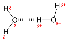
Water is a good solvent, meaning many substances dissolve in it. Polar, or charged, molecules dissolve readily in water due to water's dipolar nature. As seen in the diagram below, the slight positive charge on hydrogen atoms attracts negative solutes, while the slight negative charge on the oxygen atoms of water attracts positive ions in solutes. These polar molecules are often described as hydrophilic, meaning they are attracted to water.
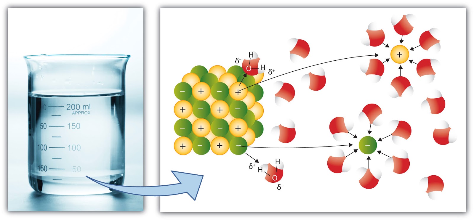
Non-polar molecules, such as lipids, cannot dissolve in water and are therefore described as hydrophobic; they are repelled by water.
The fact that so many essential polar substances dissolve in water enables them to be transported easily around animals and plants, either in the blood or xylem, to cells where they are needed inside the organism.
High Specific Heat Capacity
This means that a lot of energy is required to raise the temperature of water. Some of the heat energy is used to break the hydrogen bonds between water molecules.
This is useful to organisms because it means the temperature of water remains relatively stable, even if the surrounding temperature fluctuates significantly. Therefore, internal temperatures of plants and animals should remain relatively constant despite changes in the outside temperature. This stability is important so that enzymes do not denature or reduce in activity due to temperature fluctuations. Finally, this provides a stable environment, in terms of temperature, for aquatic organisms.
Large Latent Heat of Vaporization
This means that a lot of energy is required to convert water from its liquid state to a gaseous state. Again, this is due to the hydrogen bonds, as some energy is used to break the hydrogen bonds between water molecules to turn it into a gas.
This is advantageous to organisms because it means that water provides a significant cooling effect. For example, when humans sweat, they release water onto their skin. Large amounts of heat energy from the skin are transferred to the water to evaporate it, thus removing a lot of heat and cooling the organism.
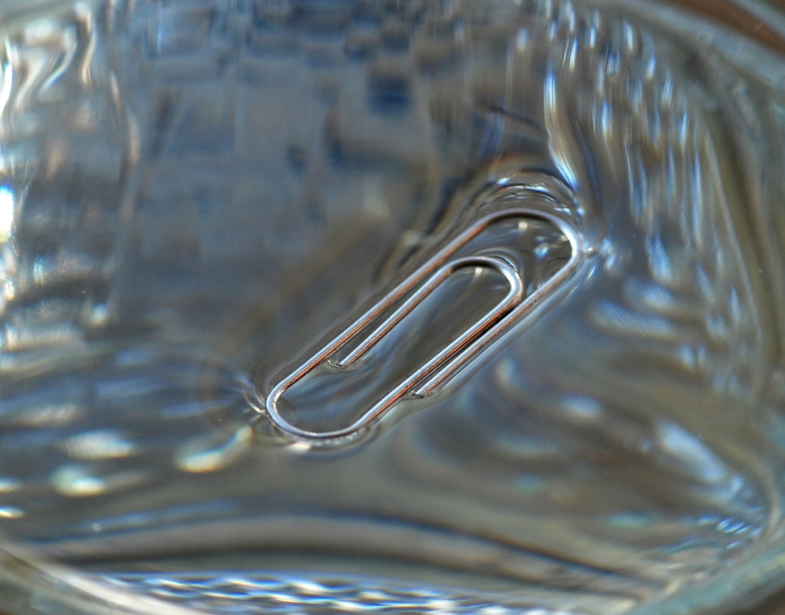
Strong Cohesion
Cohesion is the term used to describe water molecules 'sticking' together due to hydrogen bonds. Because water molecules stick together, when water moves up the xylem in plants due to transpiration, it forms a continuous column of water. This is advantageous as it is easier to draw up a column rather than individual molecules.
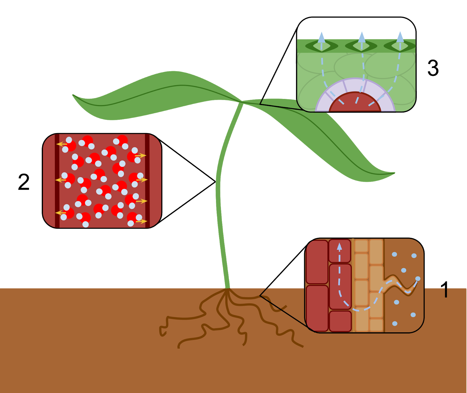
Cohesion also provides surface tension to water. This enables small invertebrates to move and live on the surface, providing them a habitat away from predators within the water. You can test this idea of surface tension by carefully placing a paperclip on water, and it should float!
- What chemical reactions have you learned already that involve water?
- Your answer should include: Hydrolysis / Condensation
- How are oxygen and hydrogen held together within a water molecule? (hint: see images above)
- Covalently
- How are water molecules held together?
- Your answer should include: Hydrogen / Bonds / Bond
- Water has an uneven distribution of electrical charges. What is the term given to describe this structural feature?
- Dipolar
- Which structural feature of water makes it a good solvent?
- Dipolarity
- Which structural feature of water provides strong cohesion, a high latent heat of vaporization, and a high specific heat capacity?
- Your answer should include: Hydrogen / Bonds / Bond
Inorganic Ions
Inorganic ions occur in the cytoplasm of organisms, some in high concentrations and others in very low concentrations. Each type of ion has a specific role, depending on its properties, and these roles are relevant to a wide range of topics across A-Level biology.
The key ions that you need to be familiar with are:
- Hydrogen and hydroxide ions impact on pH – this is relevant when considering enzymes and proteins denaturing, increasing heart rate, and the Bohr effect on hemoglobin. Hydrogen carbonate provides a source of carbon dioxide to plants when dissolved in a solution, but in human blood, it lowers the pH.
- Chloride ions and their inhibitory effect at a synapse.
- Sodium and potassium ions in the co-transport of glucose and amino acids – this is relevant in the absorption of glucose and resting/action potential in the nervous system.
- Phosphate ions as components of DNA and of ATP – this is relevant to DNA, RNA, and ATP structure.
- Ammonium ions as a result of the decay of amino acids in decomposition and deamination.
- Nitrate ions are absorbed through plant root hair cells and are essential for the creation of proteins and nucleic acids.
Lipids
Lipids are biological molecules that contain the elements carbon, hydrogen, and oxygen. These are the same elements found in carbohydrates, but unlike carbohydrates, lipids have a lot less oxygen.
Lipids are non-polar molecules or uncharged and are therefore insoluble in water. They will dissolve in organic solvents, such as ethanol. Due to the fact that they are non-polar and not soluble in water, they are described as being hydrophobic.
Lipids are made up of two molecules, fatty acids and glycerol, and they do not form polymers.
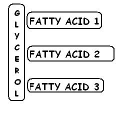
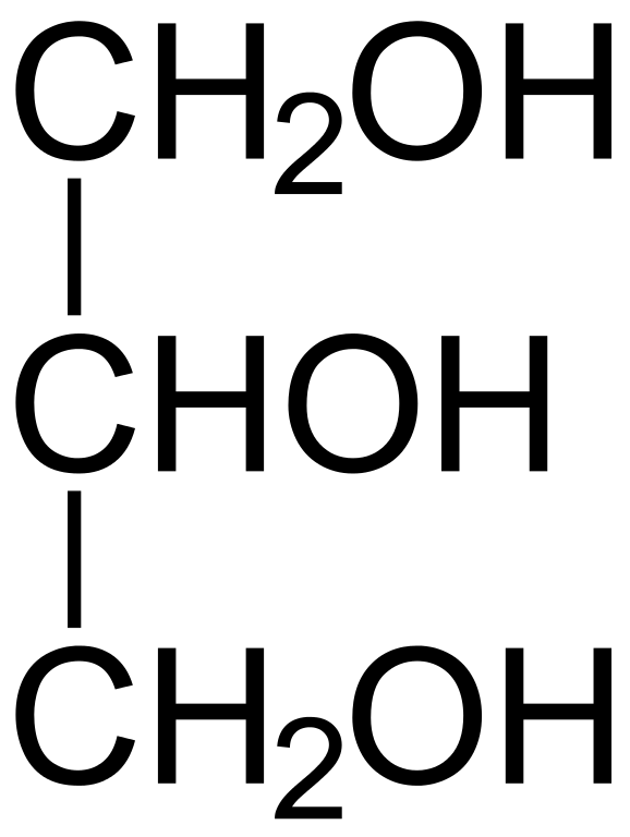
Glycerol
There are many types of lipids, but triglycerides (fats and oils) and phospholipids (in cell membranes) are the two key types you need to learn.
All lipids are made up of glycerol molecules and fatty acids.
Fatty Acids
Fatty acids are long chains of carbon and hydrogen atoms with a carboxyl group at one end (COOH). This hydrocarbon tail can vary in length.
Fatty acids can be either saturated or unsaturated, and this refers to whether there are any double bonds between the carbon atoms. Saturated Fatty Acids have no double bonds between the carbon atoms. Unsaturated Fatty Acids have at least one double bond between carbon atoms. If they have one double bond, they are described as being monounsaturated; if there are many double bonds, it is polyunsaturated.
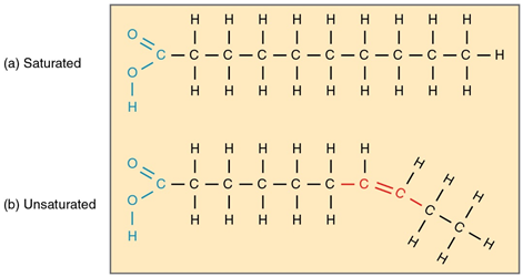
Saturated fatty acids hold as many H atoms as possible, due to the lack of double bonds - hence the name saturated. This results in a relatively straight shape, so molecules can be tightly packed in parallel. This tight structure results in these lipids being solid, or fats.
Unsaturated fatty acid chains kink where the double bonds are, and are therefore far less straight. This means the lipid molecules can't be as tightly packed and are in a liquid state, oils.
Triglycerides
Triglycerides are made up of one glycerol molecule and three (tri) fatty acids. These fatty acids are each bonded onto the glycerol by a condensation reaction. The condensation reaction occurs between the carboxyl group (COOH) of the fatty acid and the hydroxyl group (OH) of the glycerol.
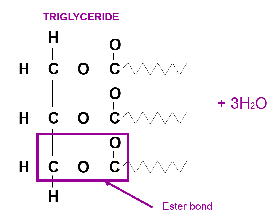
The bond that forms between the glycerol and carboxyl group of the fatty acids is an ester bond.
How the triglyceride structure results in its properties:
-
It is an energy storage. Due to the large ratio of energy-storing carbon-hydrogen bonds compared to the number of carbon atoms, a lot of energy is stored in the molecule.
-
Due to the high ratio of hydrogen to oxygen atoms, they can act as a metabolic water source. This is because triglycerides can release water if they are oxidized. This is essential for animals in the desert, such as camels.
-
As lipids are large, hydrophobic molecules, they are insoluble in water. This means they will not affect water potentials and osmosis.
-
Lipids are relatively low in mass. This means a lot can be stored in an animal without increasing its mass and preventing movement.
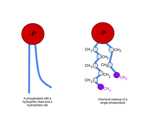
Phospholipids
Phospholipids are made up of a glycerol molecule, two fatty acid chains, and a phosphate group (attached to the glycerol). The two fatty acids also bond to the glycerol via two condensation reactions, resulting in two ester bonds.
How the phospholipid structure results in its properties.
The phosphate molecule, described as the hydrophilic 'head' of a phospholipid, can interact with water as it is charged. Due to the phosphate being charged, it repels other fats. The fatty acid chain is not charged. It is known as the hydrophobic 'tail,' and it repels water but will mix with fats.
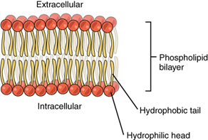
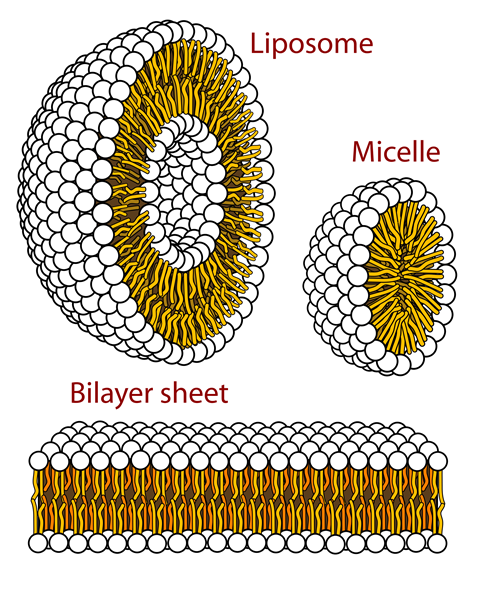
Due to these two regions on a phospholipid that act differently, it is classified as a polar molecule. The impact this has is that if phospholipids are in water, they will move to a position where the heads are exposed to water and the tails are not. This explains many of the properties stated below:
-
This behavior of the tails moving away from water results in the formation of a phospholipid bilayer membrane structure, which forms the plasma membrane around cells.
-
The hydrophilic nature of the phosphate head enables the surface of the plasma membrane to stay in place.
-
The phospholipid bilayer arrangement enables carbohydrates to attach and form important receptors on the membrane (glycolipids).
Ethanol Emulsion Test
The emulsion test is how to check for the presence of lipids:
- A few drops of the sample are added to ethanol. This is shaken to dissolve the sample in ethanol (You must say "dissolve" to get this mark).
- Then, distilled water is added.
- If a cloudy white, like milk, precipitate forms, then a lipid is present.
Proteins
Proteins are large polymers.
Protein Structure
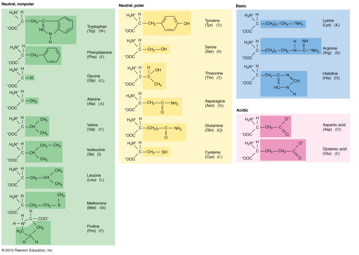
Proteins are large polymers made up of the monomer amino acids.
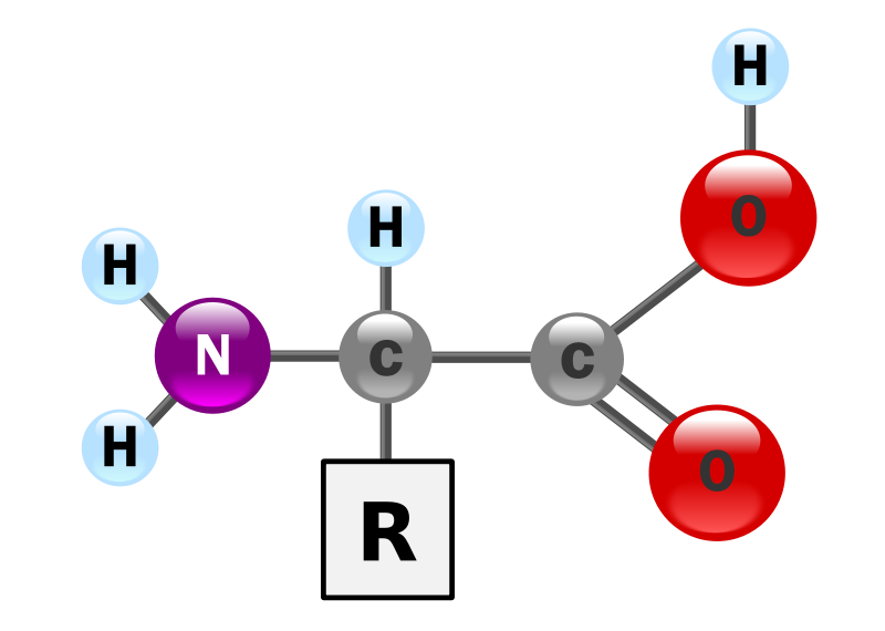
Amino acid (R is the group that is different on all 20 amino acids)
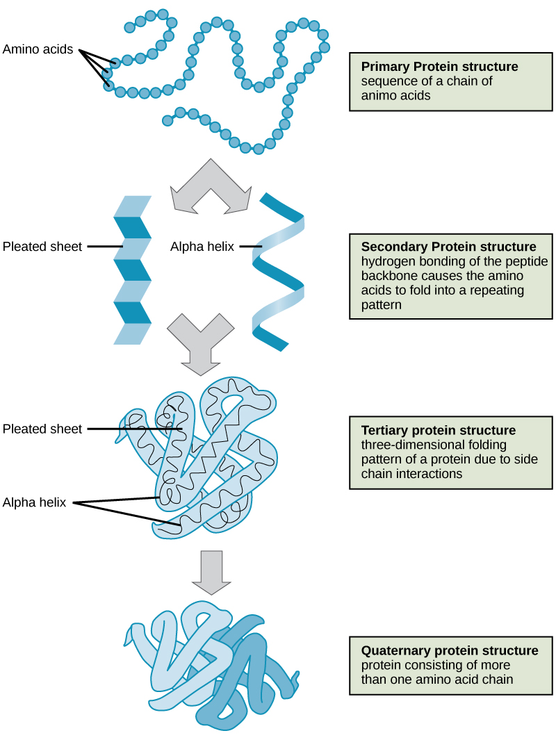
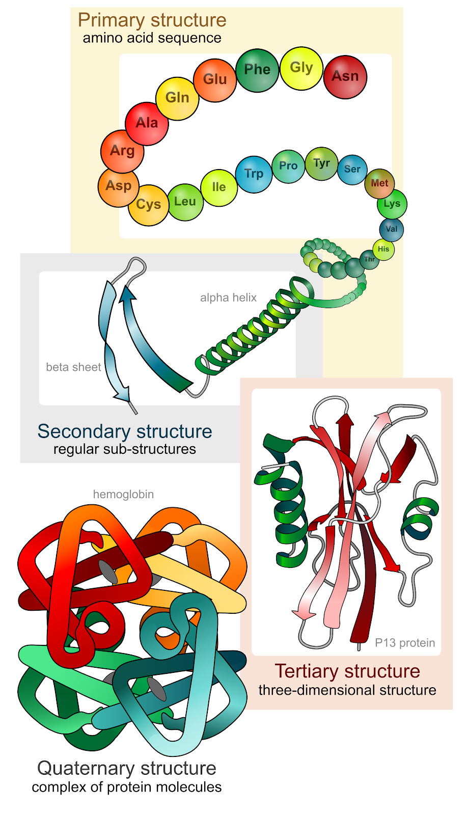
These amino acids are arranged in a series of structures to create the finished 3D protein. There are up to four levels of structural arrangements in a protein, see the image below, which will each be explained fully.
Proteins polymer chains, or polypeptides, are created on the ribosome in cells and are then further folded and modified in the Golgi apparatus.
Primary Structure
The first structure that forms in the creation of a protein is the polypeptide chain. Proteins are all made up of one or more polypeptide chains folded into highly specific 3D shapes. The exact definition of a primary structure is:
The sequence of amino acids in a polypeptide chain
This would be a one-mark question, and it is essential that you state the word ‘sequence’. The order the amino acids are bonded in is determined by DNA. This specific order of amino acids will alter how the protein further folds and is bonded, enabling unique functions.
There are 20 different amino acids that can form the primary structure. The polypeptide chain is created by a series of condensation reactions occurring between amino acids.
Secondary Structure
The sequence of amino acids causes parts of a protein molecule to bend into α helix shapes or fold into β pleated sheets.
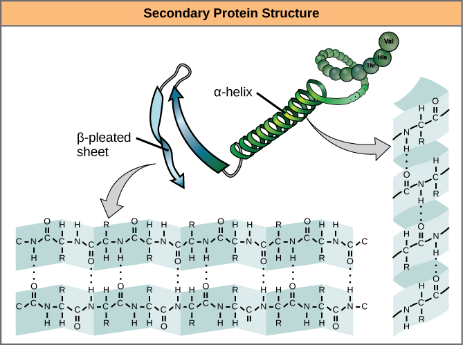
Hydrogen bonds between the carboxyl groups of one amino acid and the amino group of another are what hold the secondary structure in place. Hydrogen bonds are individually weak bonds, but when there are large numbers of them, they provide collective strength.
Describing the secondary structure would be a 2-mark question.
The two marks would be for stating:
-
Folding the primary structure into α helix shapes or fold into β pleated sheets
-
Held in place by hydrogen bonds
Tertiary Structure
The secondary structure is bent and folded to form a precise 3D shape. This unique 3D shape is held in place by hydrogen bonds, ionic bonds, and sometimes disulfide bonds. Disulfide bonds (di meaning 2) only form between the R-groups of two amino acids that contain sulfur.
Describing the tertiary structure of a protein is a 3-mark question.
The 3 marks are typically:
-
The further folding of the secondary structure
-
To create a unique 3D structure
-
Held in place by hydrogen, ionic, and disulfide bonds.
Make sure you always mention the bonds involved when describing protein structures; there are always marks for this because without these bonds, the unique shapes are not maintained.
Quaternary Structure
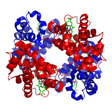
A protein that is made up of more than one polypeptide chain is a quaternary structure protein. It is still folded into a 3D shape and held by hydrogen, ionic, and disulfide bonds. Hemoglobin is the typical example given. Hemoglobin is made up of 4 polypeptide chains.
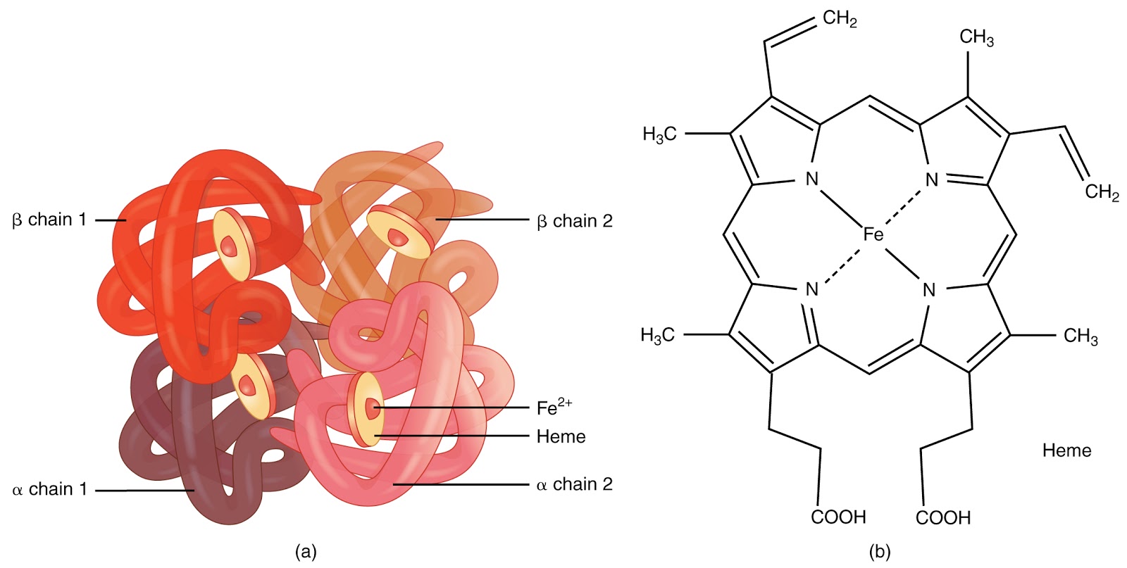
On the diagram of hemoglobin, you can also see extra molecules attached that are not part of the polypeptide chains. Any group that is attached to a protein but is not made up of amino acids is known as a prosthetic group. Iron is the prosthetic group in hemoglobin. A protein that has a prosthetic group can be described as a conjugated protein, which simply means a non-protein group is added onto it.
Fibrous and Globular Proteins
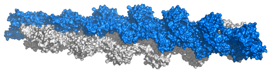
The 3D folding in tertiary and quaternary proteins generally results in either a shape that is spherical, called globular proteins, or a long rope-like shape, called fibrous proteins.
Fibrous Protein Fact File:
- Polypeptide chains form long twisted strands linked together
- Stable structure
- Insoluble in water
- Strength gives structural function
- e.g., collagen in bone and keratin in hair
Globular Protein Fact File:
- Polypeptide chains ‘roll up’ into a spherical shape
- Relatively unstable structure
- Soluble
- Metabolic functions
- e.g., all enzymes, antibodies, some hormones (e.g., insulin), hemoglobin
Cellulose
Unlike starch and glycogen, the function of cellulose is to provide structural strength in plants. Cellulose is located in the cell wall of plants and, therefore, prevents cells from bursting if they take in excess water.
Structure of Cellulose
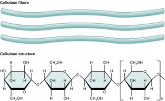
Cellulose is the only polysaccharide that is made up of β-glucose monomers. These monomers are joined by 1,4-glycosidic bonds only. For this reason, the cellulose polymer is unbranched.
These long, straight chains of β-glucose accumulate and lie parallel to each other. The parallel chains are then held together by many hydrogen bonds, and the sheer number of hydrogen bonds provides strength. This structure is called a fibril, or microfibril. Fibrils will then also align in parallel and are held in place by even more hydrogen bonds to form a cellulose fiber.
- Which polysaccharide contains 1-4 and 1-6 glycosidic bonds?
- Your answer should include: Starch / Glycogen
- Which polysaccharide is made up of the monomer β-glucose?
- Cellulose
- Which polysaccharide only forms unbranched structures?
- Cellulose
- Which is the only polysaccharide found in animal cells?
- Glycogen
- Which polysaccharide contains microfibrils?
- Cellulose
- Which polysaccharide contains amylose and amylopectin?
- Starch