Contents
Communication and Homeostasis
Communication
Cells communicate through chemical signaling, enabling communication between adjacent and distant cells. This communication involves different hormones and nervous impulses, essential for coordinating organ activities.
Neuronal Communication
The nervous system consists of the peripheral (PNS) and central (CNS) nervous systems. The PNS includes receptors, sensory and motor neurons, while the CNS encompasses coordination centers like the brain and spinal cord.
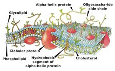
Electrical impulses in neurons result from ion movement across membranes. Therefore, understanding this topic requires knowledge of transport mechanisms across membranes (active transport, co-transport, diffusion, and facilitated diffusion) and plasma membrane structure.
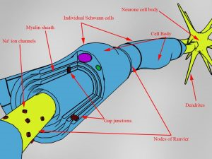
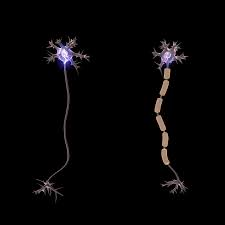
Myelinated Neuron
The structure of a myelinated motor neuron is shown below.
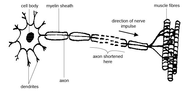
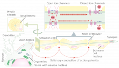
The cell body of the neuron contains organelles typical of animal cells, including the nucleus. It's in the cell body where proteins and neurotransmitter chemicals are produced.
Dendrites are branched extensions protruding from the cell body, carrying action potentials to neighboring cells.
The axon is a long, conductive fiber that carries the nervous impulse along the motor neuron.
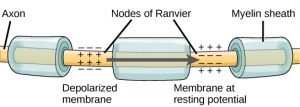
Schwann cells wrap around the axon to form the myelin sheath, a lipid layer that does not allow charged ions or impulses to pass through. There are gaps between these myelin sheaths called nodes of Ranvier.
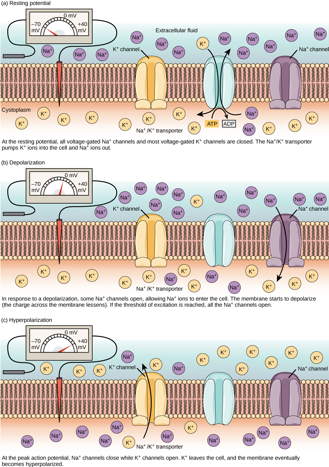
Resting Potential
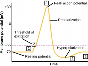
When a neuron is not conducting an impulse, there is a difference between the electrical charge inside and outside the neuron. This is known as the resting potential.
Outside the neuron, there are more positive ions (Na+) and K+ ions inside, making the inside of the neuron comparatively more negative at -70mV.
The resting potential is maintained by a sodium-potassium pump, involving active transport and ATP. The pump moves 2 K+ ions in and 3 Na+ ions out, creating a concentration gradient. As a result, K+ diffuses out and Na+ diffuses in. However, because the membrane is more permeable to K+, more K+ ions are moved out, resulting in the -70mV potential.
Action Potential
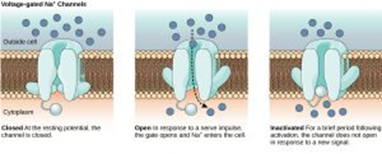
When the neuron's potential changes, it's called an action potential, resulting in a nervous impulse. Changes in membrane permeability cause depolarization and the generation of an action potential. During depolarization, Na+ continues to enter until the action potential reaches its peak, and the sodium gates close. If depolarization is not high enough to exceed a threshold, an action potential and impulse are not produced. This is called the All-or-None Principle. It ensures animals respond only to sufficiently large stimuli, preventing them from responding to minor environmental changes that could overwhelm them.
A stimulus provides energy that can open voltage-gated sodium channels in the axon membrane, causing Na+ to diffuse in, increasing the axon's positivity. This opens more voltage-gated channels, allowing even more Na+ to diffuse in. When the axon's interior reaches a threshold of +40mV, voltage-gated sodium channels close, and voltage-gated potassium ion channels open. This leads to potassium ions diffusing out, repolarizing the axon and temporarily making it more negative than -70mV, resulting in hyperpolarization. The potassium ion gates eventually close, and the sodium-potassium pump restores the resting potential.
Once an action potential is generated, it moves along the axon like a Mexican wave.
This occurs along the entire axon in non-myelinated neurons, whereas it is much quicker in myelinated axons. Action potentials can only occur at the nodes of Ranvier, where there is no myelin insulation. Therefore, localized action potentials jump from node to node, traveling much faster. This phenomenon is known as saltatory conduction.
In addition to myelination and saltatory conduction increasing the speed of conduction, temperature and axon diameter also have an impact. Higher temperature speeds up ion diffusion, increasing conduction speed. A wider axon diameter accelerates conduction because it reduces the likelihood of ions leaking across the membrane and affecting the potential.
Refractory Period
Immediately after an action potential is generated, the membrane enters a refractory period during which it can't be stimulated because sodium channels are recovering and cannot be opened. This means that the voltage-gated sodium channels are closed. This is important for several reasons:
Firstly, it ensures the production of discrete impulses, preventing an action potential from being generated immediately after another, ensuring they remain separate.
Secondly, it ensures that action potentials travel in one direction, preventing them from spreading out in two directions and preventing a response.
Finally, it limits the number of impulse transmissions, preventing overreaction to a stimulus and overwhelming the senses.
- What is the term given to the potential when the neuron is at rest?
- Resting
- What is the term given to the potential when a stimulus is detected?
- Action
- What is the term given to the period after a stimulus generates a response?
- Refractory period
- What determines whether the sodium channels within axon membranes open or close?
- Voltage
- What is the name of the insulating layer that wraps around an axon?
- Myelin Sheath
- Where in a motor neuron is the nucleus found?
- Cell Body
Homeostasis
Homeostasis is the maintenance of a constant internal environment via physiological control systems. These control systems keep temperature, blood pH, blood glucose and water potential within set limits.
Importance of Homeostasis
If body temperature is too low there will be insufficient kinetic energy for enzyme-controlled reactions, and if body temperature is too high then enzymes will denature. Either way, metabolic reactions could slow to the point that cells die. Alterations in blood pH will also result in enzymes denaturing.
Glucose is needed for respiration, so a lack of glucose in the blood could result in cell death. If blood glucose levels are too high, then this will lower the water potential of the blood and water will leave surrounding cells by osmosis and prevent normal cell function. If the water potential of the blood is too low, water will move into cells by osmosis and can cause them to burst.
Negative Feedback
Negative feedback is how a deviation from the normal values are restored systems to their original level. This involves the nervous system and often hormones too, as shown in the flow diagram.
Positive Feedback
Positive feedback is when a deviation from the normal values triggers a response to increase the deviation further, for example during childbirth.
Excretion as an Example of Homeostatic Control
Mammalian Liver
The liver is essential for glycogen storage, detoxification and in the formation of urea. The liver contains a range of different enzymes that make these processes possible.
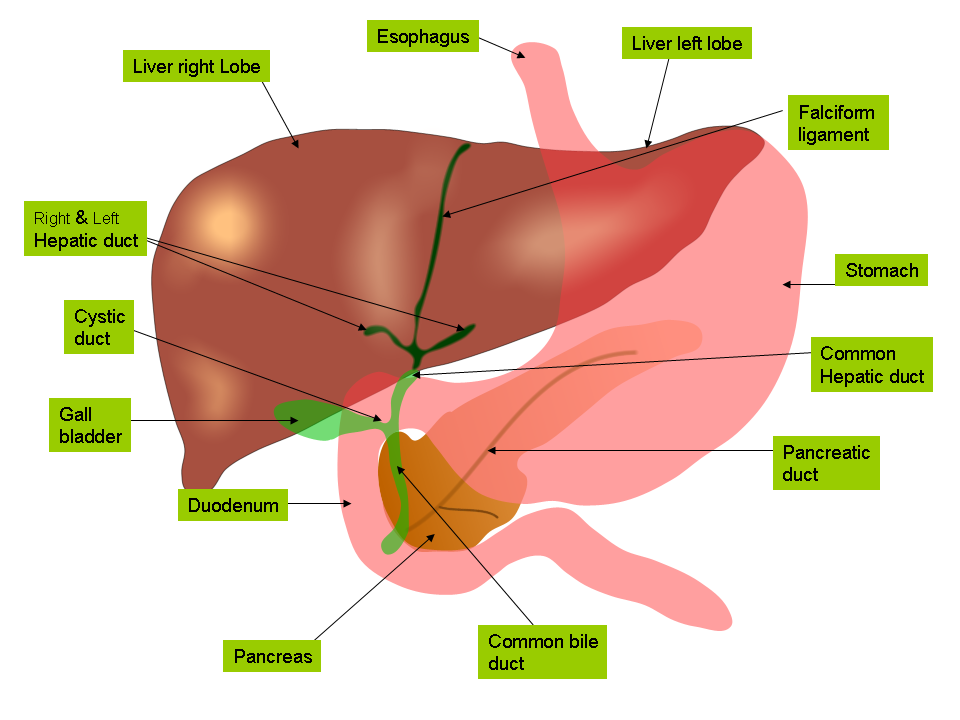
The ornithine cycle (urea cycle) is how urea is produced from ammonia, ready to be transported to the kidneys and excreted. Excess proteins are deaminated in the liver (broken down to amino acids), which are then converted into ammonia. Ammonia is highly toxic, which is why it is converted to urea before being transported in the blood.
Detoxification is the neutralisation/breakdown of unwanted chemicals such as alcohol, drugs, hormones and toxins produced in chemical reactions in the body.
Mammalian Kidney and Osmoregulation
This is the process of controlling the water potential of the blood.
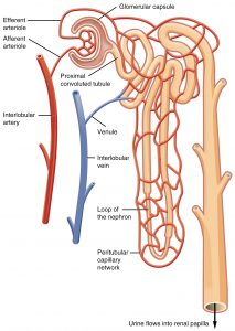
The nephron is structure in the kidney where the blood is filtered, and useful substances are reabsorbed into the blood. This is shown in the diagram below.
Thermoregulation
Endotherms are able to regulate their own body temperature through a nervous response. Peripheral receptors in the skin detect a change in the external temperature. This sends an impulse along a sensory neurone to the brain, where the hypothalamus coordinates the impulse. This will trigger a response, which if you are too hot could be for glands to produce more sweat, vasodilation or behavioural (e.g. move to the shade, remove clothes, fan yourself). If you are too cold the responses are for glands to produce less sweat, muscles to contract and relax (shivering) to generate heat and behavioural (wear more clothes).
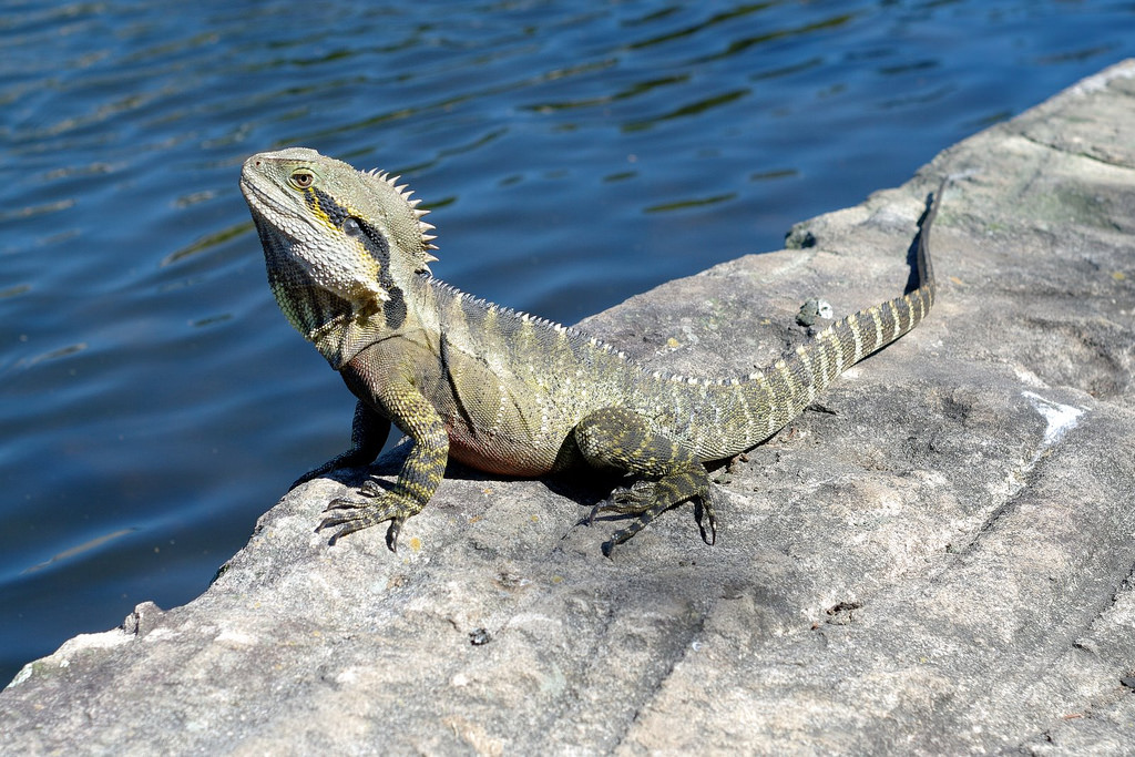
Ectotherms cannot regulate their internal temperature and can only control it through their behaviour. This is why cold blooded animals, ectotherms, often bask on hot rocks to absorb the radiating heat.
The graph shows how endotherms, the mouse, are able to regulate their temperature so there is little fluctuation even when the external temperature increases: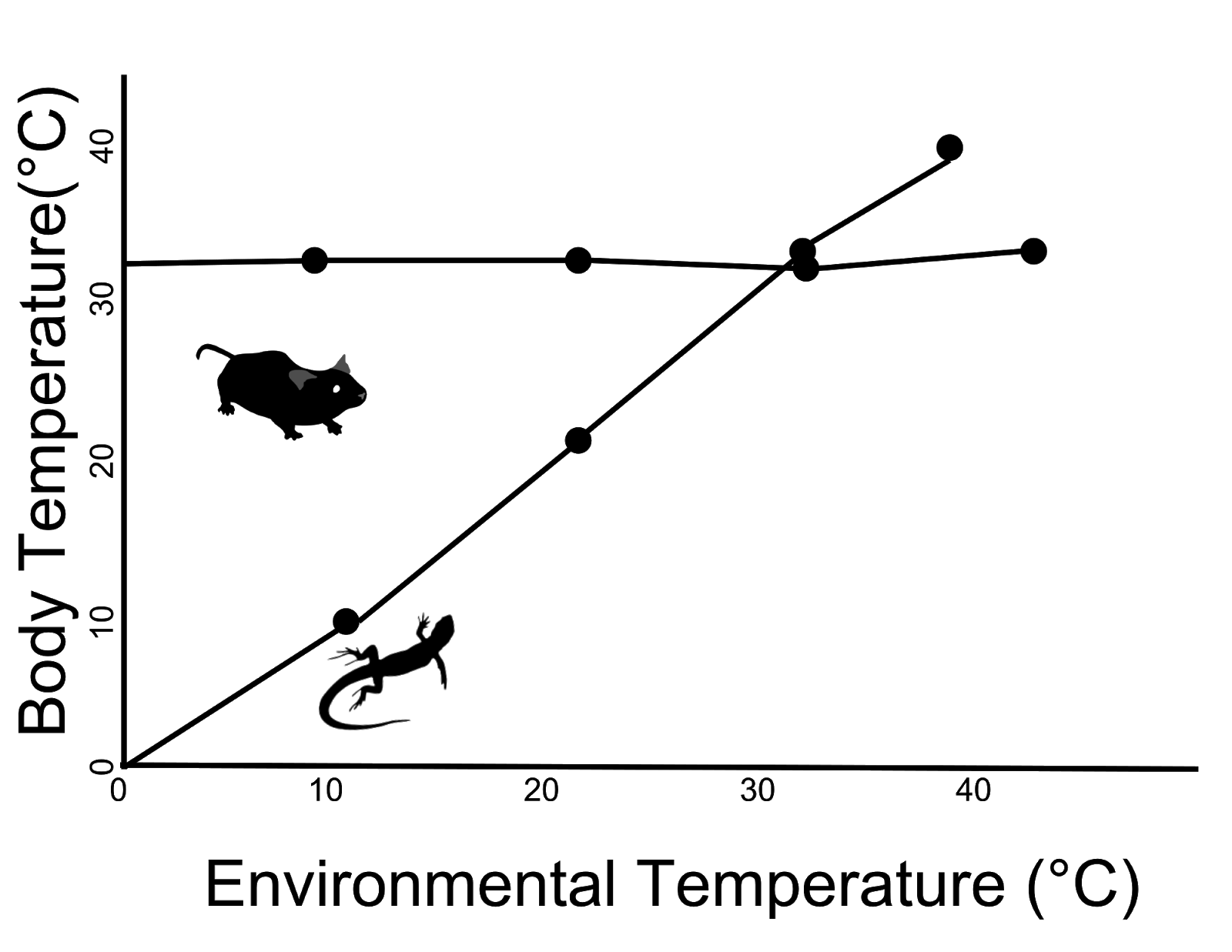
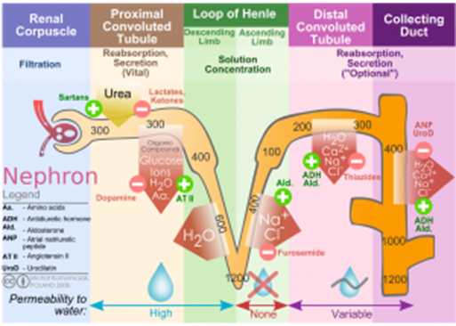
Nephron Structure
- The Bowman’s (renal) capsule - Ultrafiltration occurs here because the afferent arteriole (entering the glomerulus) is wider than the efferent arteriole (leaving the glomerulus) creating high hydrostatic pressure. Small molecules and water are forced out of the capillaries into the renal capsule, and large proteins and blood cells remain in the blood.
- Proximal convoluted tubule - The walls are made of microvilli epithelial cells to provide a large surface area for diffusion of glucose into the cells from the PCT. Glucose is then actively transported out of the cells into the intercellular space to create a concentration gradient. Glucose can then diffuse into the blood again.
- Loop of Henle - Sodium ions are actively transported out of the ascending limb into the medulla to create a low water potential. Water moves out of the descending limb and out of the distal convoluted tubule and collecting duct by osmosis due to this water potential gradient.
The liquid remaining in the collecting duct forms urine. It contains water, dissolved salts, urea, and other substances such as hormones.
More or less water can be reabsorbed at the collecting duct, depending on the water potential of the blood. The hypothalamus in the brain monitors the water potential of the blood. If the water potential of the blood decreases, water will move out of the osmoreceptor cells by osmosis, resulting in the cells shriveling. This triggers other cells in the hypothalamus, which send a signal to the posterior pituitary gland. The posterior pituitary will then release antidiuretic hormone (ADH) into the blood.
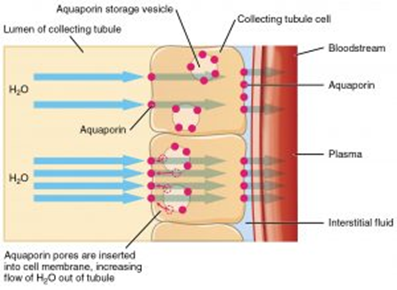
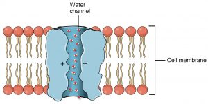
ADH will bind to complementary receptors located only on target cells in the DCT and collecting duct. When it binds, it activates adenyl cyclase to make cAMP. This activates an enzyme that causes vesicles containing aquaporins to fuse with the membrane. Aquaporins and channel proteins allow water to transport across the membrane. As a result, the membrane becomes more permeable to water, and more will leave to be reabsorbed back into the blood.
If kidney failure occurs, then the blood is not filtered properly, which can lead to a buildup of urea in the blood and an electrolyte imbalance (mineral ions). Two potential treatments for this are dialysis: a machine that the patient’s blood is passed through 3 to 4 times per week, which performs the function of a kidney to filter the blood. A second option is a kidney transplant, but this depends on the availability of a kidney from a donor that is a match.
Urine in Diagnosis
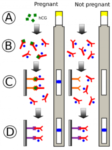
Due to urine being composed of substances filtered out of the blood, it can be used to test for diabetes, pregnancy, anabolic steroids, and drugs.
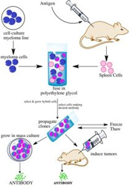
A pregnancy test uses monoclonal antibodies to detect the presence of the human growth hormone, which is produced by pregnant women. A monoclonal antibody is a single type of antibody that can be isolated and cloned. This is used for targeting medication to specific cell types by attaching a therapeutic drug to an antibody and medical diagnosis. Creating monoclonal antibodies requires mice to produce the antibodies and tumor cells, which leads to ethical debates as to whether this use of animals is justified to enable the better treatment of cancers in humans and to detect disease.
Synapse and Neuromuscular Junction
Synapses are the gaps between the end of the axon of one neuron and the dendrite of another one. Here, the action potential is transmitted as neurotransmitters that diffuse across the synapse.
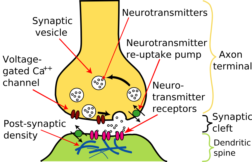
Neuromuscular junction
This is a synapse that occurs between a motor neuron and a muscle and is very similar to a synaptic junction.
| Neuromuscular Junction | Cholinergic Synapse |
| Unidirectional due to the neurotransmitter receptors only being on the postsynaptic membrane | |
| Only excitatory | Could be excitatory or inhibitory. Inhibitory synapses cause chloride ions to move into the postsynaptic neuron and potassium ions to move out. The combined effect of negative ions moving in and positive ions moving out makes the membrane potential increase to -80mV, hyperpolarization, and therefore an action potential is highly unlikely. |
| Connects motor neuron to muscles | Connect two neurons, which could be sensory, relay, or motor. |
| This is the endpoint for the action potential | A new action potential is generated in the next neuron. |
| Acetylcholine binds to receptors on muscle fiber membranes | Acetylcholine binds to receptors on the postsynaptic membrane of a neuron. |
Summation
Summation is the rapid buildup of neurotransmitters in the synapse to help generate an action potential by two methods: spatial or temporal summation. This is needed because some action potentials do not result in sufficient concentrations of neurotransmitter being released to generate a new action potential.
Spatial summation: many different neurons collectively trigger a new action potential by combining the neurotransmitter they release to exceed the threshold value.
Temporal summation: One neuron releases neurotransmitter repeatedly over a short period of time to add up to enough to exceed the threshold value.
Hormonal Communication
Blood Glucose Control
Blood glucose will increase following ingestion of food or drink containing carbohydrates and will fall following exercise or if you have not eaten.
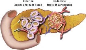
The pancreas detects changes in the blood glucose levels and contains endocrine cells in the Islets of Langerhans which release the hormones insulin and glucagon to bring blood glucose levels back to normal. Adrenaline is released by adrenal glands if your body anticipates danger and results in more glucose being released from stores of glycogen in the liver.
Key Terms:
- Glycogenesis (genesis means to make)
This is the process of when excess glucose is converted to glycogen in blood glucose is higher than normal. This occurs in the liver, and the liver can store 75-100g of glycogen. This would last you 12 hours at rest if you do not eat.
- Glycogenolysis (lysis means to breakdown)
This is the breakdown of glycogen back into glucose in the liver. This occurs when blood glucose is lower than normal.
- Gluconeogenesis (Amino acids to glucose)
This is the process of creating glucose from non-carbohydrate stores in the liver. This will occur if all glycogen has already been hydrolyzed back into glucose and your body still needs more glucose.
The above processes are controlled by the three hormones insulin, adrenaline, and glucagon.
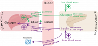
The Action of Insulin
Beta cells in the Islets of Langerhans detect if blood glucose is too high and will secrete insulin. Insulin will decrease blood glucose in the following ways:
Attaching to receptors on the surfaces of target cells. This changes the tertiary structure of the channel proteins resulting in more glucose being absorbed by facilitated diffusion.
More protein carriers are incorporated into cell membranes so that more glucose is absorbed from the blood into cells.
Activating enzymes involved in the conversion of glucose to glycogen. This results in glycogenesis in the liver.
The Action of Glucagon:
Attaching to receptors on the surfaces of target cells. When glucagon binds, it causes a protein to be activated into adenylate cyclase and to convert ATP into a molecule called Cyclic AMP (cAMP). cAMP activates an enzyme, protein kinase, that can hydrolyze glycogen into glucose.
Activating enzymes involved in the conversion of glycerol and amino acids into glucose.
The Role of Adrenaline by:
Adrenaline attaches to receptors on the surfaces of target cells. This causes a protein (G protein) to be activated and to convert ATP into cAMP. cAMP activates an enzyme that can hydrolyze glycogen into glucose. This is known as the second messenger model of adrenaline and glucagon action because the process results in the formation of cAMP, which acts as a second messenger.
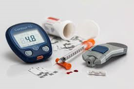
Diabetes
Diabetes is a condition where blood glucose levels cannot be properly controlled.
Type I diabetes results from the body's inability to produce insulin. It typically starts in childhood and can be the result of an autoimmune disease where the beta cells are attacked. Treatment involves insulin injections.
Type II diabetes occurs when receptors on the target cells lose their responsiveness to insulin. It usually develops in adults due to obesity and poor diet. It can be managed by regulating carbohydrate intake, increasing exercise, and sometimes using insulin injections.
The Human Brain
The human brain consists of billions of neurons and plays a critical role in coordinating responses within the body.
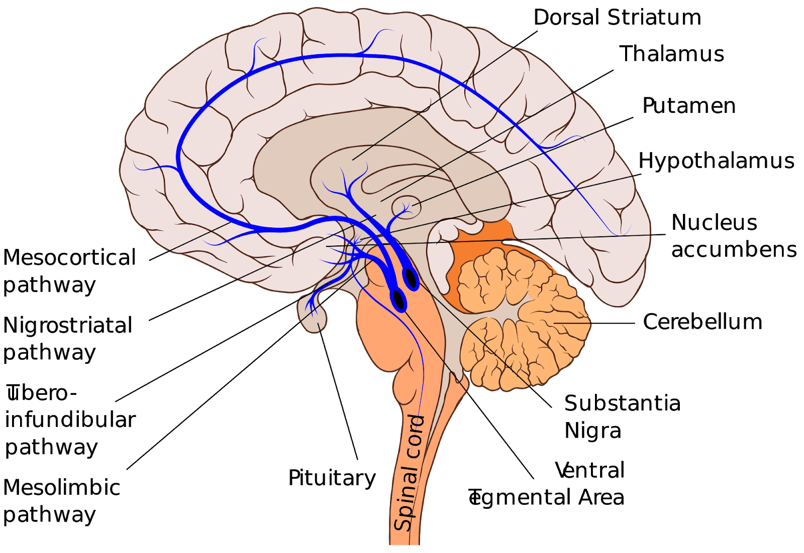
The key structures of the brain include the cerebrum, cerebellum, medulla oblongata, hypothalamus, and the pituitary gland.
Cerebrum - The largest part of the brain, shown in two shades of grey above. The lighter grey is the outer layer, known as the cerebral cortex. It is highly folded and divided into two hemispheres. Its functions encompass controlling conscious thoughts, language, intelligence, personality, high-level functions, and memory.
Cerebellum - Shown in orange above, resembling a mini cauliflower, it coordinates movement, muscles, and balance.
Medulla Oblongata - The orange structure labeled above the spinal cord. It serves as the control center for unconscious activities such as breathing and heart rate.
Hypothalamus - This small part of the brain is responsible for maintaining homeostasis, including temperature and water balance.
Pituitary Gland - The small lobed grey structure in the diagram above, known as the master gland, releases various hormones to coordinate several bodily responses, such as the oestrous cycle and osmoregulation.
Control of the Heart
Cardiac muscle is myogenic, meaning it can contract on its own, but the rate of contraction is controlled by a wave of electrical activity. The sinoatrial node (SAN) is located in the right atrium and acts as the heart's pacemaker. It initiates a wave of depolarization across the atria, causing them to contract. The atrioventricular node (AVN) is positioned near the border of the right and left ventricle within the atria.
The medulla oblongata in the brain governs heart rate via the autonomic nervous system. It consists of two parts: one linked to the sinoatrial node that increases heart rate via the sympathetic nervous system and another that decreases heart rate via the parasympathetic nervous system.
The heart rate can change in response to pH and blood pressure, and these stimuli are detected by chemoreceptors and pressure receptors in the aorta and carotid artery.
- Why do hormones only affect their target cells and not other cells?
- Hormones affect their target cells because of the specific receptors on the cell surface membrane that are complementary to the hormones.
- Why is it harmful if there is too much glucose in the blood?
- Excess glucose in the blood can lead to a reduction in water potential, causing osmosis and potential damage to cells.
- Why is it harmful if there is not enough glucose in the blood?
- Insufficient glucose in the blood can impair respiration and energy production in cells.
- Where is ADH released from?
- ADH is released from the pituitary gland.
- What does ADH stand for?
- ADH stands for Anti-Diuretic Hormone.
- What effect does ADH have on the collecting duct?
- ADH makes the collecting duct more permeable to water.
Plant and Animal Responses
Responses in Flowering Plants
Tropism is the term given when plants respond to stimuli through growth. Tropisms can be either positive (growing toward a stimulus) or negative (growing away from a stimulus). Plants respond to light, gravity, and water.

Tropisms are controlled by specific growth factors, and one key example is indoleacetic acid (IAA). IAA is a type of auxin that can control cell elongation.
Phototropisms
Shoots
Plants need light for photosynthesis, which is why they grow and bend toward light. This is controlled by IAA, resulting in positive phototropism.
Shoot tip cells produce IAA, causing cell elongation in shoots, which then diffuses to other cells. If there is unilateral light, IAA will diffuse toward the shaded side of the shoot, leading to a higher concentration of IAA there. This causes the cells on the shaded side to elongate more, making the plant bend toward the light source.
Roots
Roots do not photosynthesize and do not require light. They are better at anchoring the plant when deep in the soil, away from light. In roots, a high concentration of IAA inhibits cell elongation, causing root cells to elongate more on the lighter side, resulting in the root bending away from light. This is negative phototropism.
Gravitropism
Shoots
IAA moves from the upper side to the lower side of a shoot. In a vertical plant, this causes plant cells to elongate and grow upward. If the plant is on its side, it will cause the shoot to bend upward. This is negative gravitropism.

Roots
IAA moves to the lower side of roots so that the upper side elongates and the root bends down toward gravity, anchoring the plant. This is positive gravitropism.
Gibberellins
Gibberellins are plant hormones that help initiate germination in seeds and promote stem growth in already germinated plants.
Due to their effects, both auxins and gibberellins are used commercially to promote seed germination and plant root and shoot growth.
Responses and Reflexes
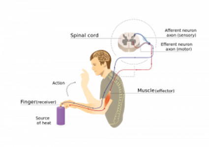
A stimulus is a detectable change in the environment. These changes can be detected by cells called receptors, triggering a response through the flow chart shown below. The effector is the structure responsible for the response, typically muscles or glands.
Stimulus → Receptor → Coordinator → Effector → Response
Organisms increase their chance of survival by responding to stimuli through different response mechanisms.
Simple Reflex
In mammals, the simplest response to a stimulus is a reflex. This is a rapid, involuntary response to danger that bypasses the brain. It is rapid because it only involves three neurons and has few synapses. Synapses slow down responses as electrical energy is converted to chemicals that diffuse across the synapse. Because no decision is involved, the response is also rapid and prevents the brain from being overloaded with situations to decide upon. Once the stimulus is detected by the receptor, an impulse is passed along the sensory neuron to a relay neuron, located in the spine. The relay neuron passes the impulse onto a motor neuron, which is connected to an effector. For example, if the stimulus is a hot object, the effector would be the muscles in your hand and arm, and the response would be their contraction to move your hand away from the dangerous hot object.
Muscles
Muscles act in antagonistic pairs against an incompressible skeleton to create movement. This can be automatic as part of a reflex response or controlled by conscious thought.
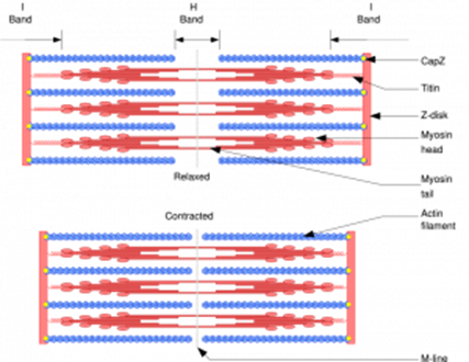
Muscle fibers are made up of millions of myofibrils, collectively providing the force for movement. Myofibrils are made up of fused cells that share nuclei and cytoplasm, known as sarcoplasm, and there is a high number of mitochondria. Myofibrils consist of two key types of protein, myosin and actin, which form a sarcomere.
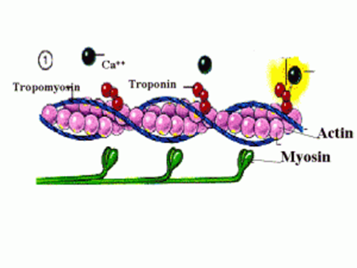
Sliding Filament Theory
When an action potential reaches a muscle, it stimulates a response. Calcium ions enter and cause the protein tropomyosin, which blocks binding sites for the myosin head of the actin, to move and uncover the binding sites. While ADP is attached to the myosin head, it can bind to the binding site on the actin to form a cross-bridge. The angle created in this cross-bridge creates tension, and as a result, the actin filament is pulled and slides along the myosin. In doing so, the ADP molecule is released. An ATP molecule can then bind to the myosin head and causes it to change shape slightly, detaching it from the actin. Within the sarcoplasm, there is the enzyme ATPase, which is activated by the calcium ions, to hydrolyze the ATP on the myosin head into ADP, releasing enough energy for the myosin head to return to its original position. This entire process repeats continually while the calcium ions remain high, and therefore while the muscle remains stimulated by the nervous system.
Active muscles therefore require a high concentration of ATP. In times when aerobic respiration cannot create enough ATP to meet this demand, anaerobic respiration occurs. The chemical phosphocreatine, which is stored in muscles, assists this by providing phosphate to regenerate ATP from ADP.
Slow and Fast Skeletal Muscle Fibers
| Slow-twitch fibers | Fast-twitch fibers | |
| Structure | Contain a large store of myoglobin, a rich blood supply, and many mitochondria. | Thicker and have more myosin filaments, a large store of glycogen, a store of phosphocreatine to help make ATP from ADP, and a high concentration of enzymes involved in anaerobic respiration. |
| Location | Calf muscles | Biceps |
| General properties | Contract slower and can respire aerobically for longer periods due to the rich blood supply and myoglobin oxygen store. These muscles are adapted for endurance work, like marathons. | Contract faster to provide short bursts of powerful contraction. These are adapted for intense exercise, such as sprinting or weightlifting. |