Contents
Transport in Animals
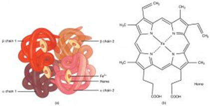
Hemoglobin
Hemoglobin is a protein with a quaternary structure, briefly mentioned in Topic 1. Hemoglobins are a group of proteins and different forms are found in different organisms. The role of hemoglobin and red blood cells is to transport oxygen.
The loading, transport, and unloading of oxygen varies in different conditions and for the different forms of hemoglobin, and this can be presented on an oxyhemoglobin dissociation curve.
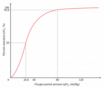
The affinity hemoglobin has for oxygen changes depending on how many oxygen molecules are already associated with it. Hemoglobin can associate with, or load, four oxygen molecules and as each molecule binds the shape of hemoglobin changes making the binding of further oxygen molecules easier. Therefore, in areas with a high partial pressure of oxygen, meaning a high concentration, the affinity of hemoglobin for oxygen is high and it loads more oxygen. In humans, the alveoli have a high partial pressure of oxygen and therefore hemoglobin will readily load with oxygen here.
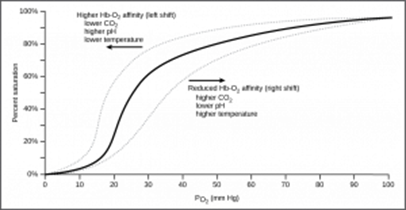
A high partial pressure of carbon dioxide results in a reduced affinity for oxygen and the hemoglobin will more readily unload oxygen, this is known as the Bohr effect. When carbon dioxide dissolves in liquid carbonic acid forms, and this decrease in pH changes the shape of hemoglobin slightly, which is why the affinity for oxygen decreases. In respiring tissues, there will be a high partial pressure of carbon dioxide and therefore the oxygen dissociates from the hemoglobin. This is advantageous, as the hemoglobin delivers the oxygen to the site of respiring cells so that aerobic respiration can continue.
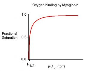
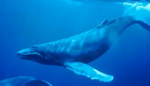
Many animals are adapted to their environment by possessing different types of hemoglobin with different oxygen transport properties. Animals like lugworms, whales, and even human fetuses have myoglobin. Myoglobin has a very high affinity for oxygen, even at very low partial pressures. Therefore, it acts like an oxygen store, holding on to oxygen and not dissociating until nearly all oxygen has been used up in cells.
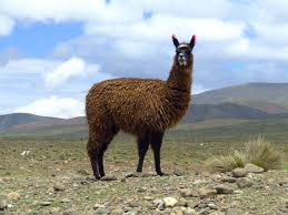
A different example is llamas and other animals at high altitude. The atmospheric pressure is low and so there is a lower partial pressure of oxygen. Llamas have a type of hemoglobin with a higher affinity of oxygen so that despite the low partial pressure of oxygen, it is still loaded onto hemoglobin.
Circulatory System in Mammals
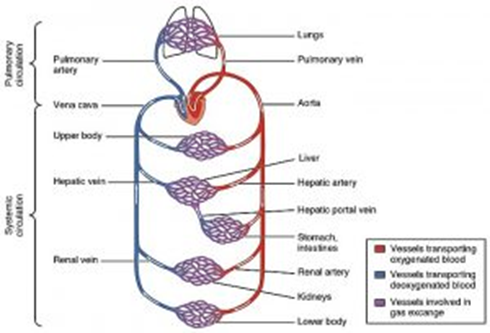
You need to have knowledge of the coronary arteries, the blood vessels entering and leaving the heart, lungs, and kidneys. Some of these can be seen below in the diagram of the mammalian double circulatory system.
Human Heart
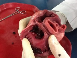
The diagram shows the key structures of the human heart. You need to know how to dissect the heart. You would need to wear gloves for hygiene and use a sharp scalpel to cut the cardiac tissue to then see the inside of the heart, such as the valves and tendons.
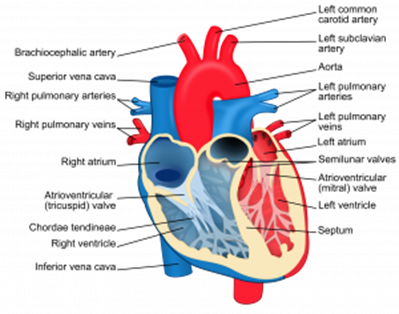
The left ventricle has the thickest muscular wall so that it can contract with more force to pump blood at higher pressure so that it will flow all the way around the body. The right ventricle only pumps blood to the lungs, which is much closer and requires blood to flow slower to allow time for gas exchange, so the muscular wall is much thinner. The atria both have very thin muscular walls, as they only pump blood to the ventricles.
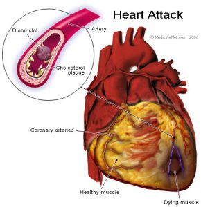
The coronary arteries supply the cardiac muscle with oxygenated blood. Therefore, a clot in a coronary artery will prevent the oxygen supply to cardiac muscle and result in anaerobic respiration and eventually the muscle won't be able to contract and relax- this leads to myocardial infarction (a heart attack).
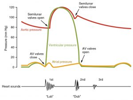
The cardiac cycle is a series of muscle contractions and relaxation resulting in pressure and volume changes that cause valve movements and maintain a unidirectional flow of blood. Valves open when there is a higher pressure behind them.
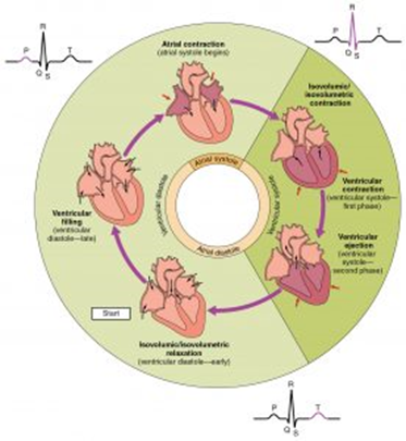
Atrial systole is when the atria contract. This increases the pressure in the atria and forces the atrioventricular valves to open and blood flows into the ventricles. Ventricular systole is when the ventricles contract and the atria are relaxing. This increases the pressure in the ventricles, causing the atrioventricular valves to shut and the semilunar valves to open. Therefore, blood flows out of the ventricles and into the pulmonary artery and aorta.
You can calculate the cardiac output using the formula:
CO = stroke volume × heart rate
Heart rate is how many beats per minute. Stroke volume is the volume of blood pumped in one beat.
Control of the Heart
Cardiac muscle is myogenic, but the rate of contraction is controlled by a wave of electrical activity.
The sinoatrial node (SAN) is in the right atrium and is known as the pacemaker. The SAN will release a wave of depolarization across the atria, causing it to contract. The atrioventricular node (AVN) is located near the border of the right and left ventricle within the atria still.
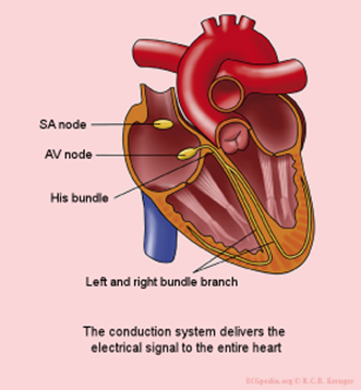
The AVN will release another wave of depolarization when the first reaches it. There is a non-conductive layer between the atria and ventricles which prevents the wave of depolarization traveling down to the ventricles. Instead, the bundle of His, running through the septum can conduct and transmit the wave of depolarization down the septum and the Purkinje fibers in the walls of the ventricles. As a result, the apex and then walls of the ventricles contract. There is a short delay before this happens, while the AVN transmits the second waves of depolarization. This allows enough time for the atria to pump all the blood into the ventricles. Finally, the cells repolarize, and the cardiac muscle relaxes.
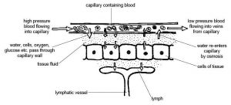
Tissue Fluid
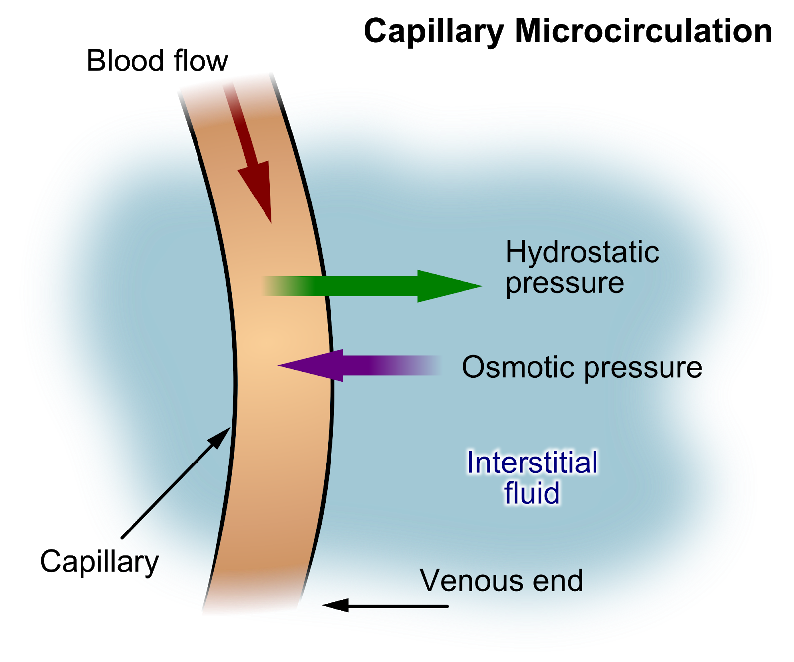
Capillaries have small gaps in the walls so that liquid and small molecules can be forced out, this forms tissue fluid.
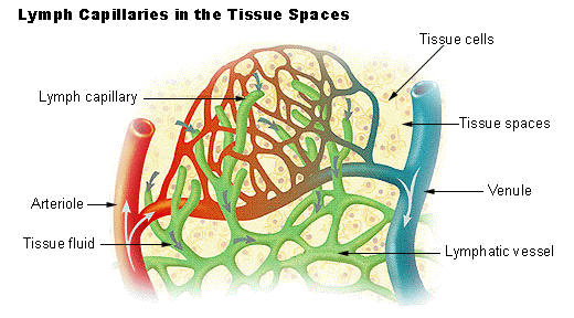
As blood enters the capillaries from arterioles, the smaller diameter results in a high hydrostatic pressure so water, glucose, amino acids, fatty acids, ions, and oxygen are forced out. This bathes the cells in substances they may need.
Large molecules remain in the capillaries and therefore create a lowered water potential. Towards the venule end of the capillaries, the hydrostatic pressure is lowered due to the loss of liquid, but the water potential is very low.
As a result, the water re-enters the capillaries by osmosis. Not all the liquid will be absorbed, as equilibrium will be reached. The rest of the tissue fluid is absorbed into the lymphatic system and eventually drains back into the bloodstream near the heart.
- Why is it an advantage that arteries have thick elastic walls?
- Your answer should include: Withstand / High / Pressure
- What substances will be found in tissue fluid?
- Your answer should include: Water / Small / Molecules
- What are the four major blood vessels associated with the heart?
- Your answer should include: Vena / Cavae / Aorta / Pulmonary / Vein / Artery
- What is the type of hemoglobin that has a very high affinity for oxygen?
- Myoglobin
- What is the term given to when carbon dioxide lowers the affinity of hemoglobin for oxygen?
- Your answer should include: Bohr / Effect
- What is the name of the human pacemaker?
- Your answer should include: SAN / Sinoatrial / Node
Blood Vessels
Arteries carry blood Away (hint to remember -A Away) from the heart and into arterioles. The arterioles are smaller than arteries and connect to the capillaries. The capillaries connect the arterioles to the veins. The veins carry blood back into the heart (hint to remember- veINs carry blood IN).
| Property | Arteries | Arterioles | Veins | Capillaries | |||
| Muscle layer | Thicker than veins so that constriction and dilation can occur to control volume of blood. | Thicker than in arteries to help restrict blood flow into the capillaries. | Relatively thin so it cannot control the blood flow. | No muscle layer. | |||
| Elastic layer | Thicker than veins to help maintain blood pressure. The walls can stretch and recoil in response to the heartbeat. | Thinner than in arteries as the pressure is lower | Relatively thinner as the pressure is much lower. | No elastic layer | |||
| Wall thickness | Thicker wall than veins to help prevent the vessels bursting due to the high pressure. | Thinner as pressure is slightly lower. | Thin as the pressure is much lower so there is a low risk of bursting. The thinness means the vessels are easily flattened, which helps the flow of blood up to the heart. | One cell thick consisting of only a lining layer. This provides a short diffusion distance for exchanging materials between the blood and cells. | |||
| Valves | No | No | Yes | No |
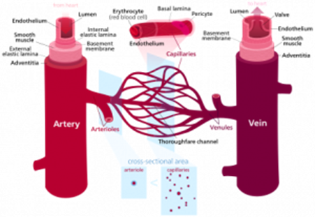
Capillaries form capillary beds as exchange surfaces, which are many branched capillaries. These all have a narrow diameter to slow blood flow. Red blood cells can only just fit through and are squashed against the walls, and this maximizes diffusion.