Contents
Nervous System
Nervous System
The nervous system is made up of the peripheral and central nervous systems. The PNS includes the receptors, sensory and motor neurons, while the CNS consists of the coordination centers such as the brain and spine.
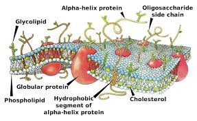
The electrical impulses that pass along neurons are due to the movement of ions across membranes. Therefore, understanding transport across a membrane (active transport, co-transport, diffusion, and facilitated diffusion) and plasma membrane structure is key to understanding this topic.
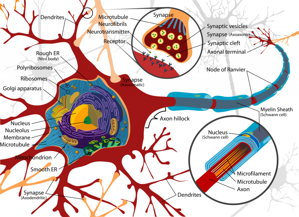
Myelinated Neuron
The structure of a myelinated motor neuron can be seen below:
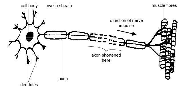
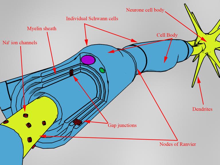
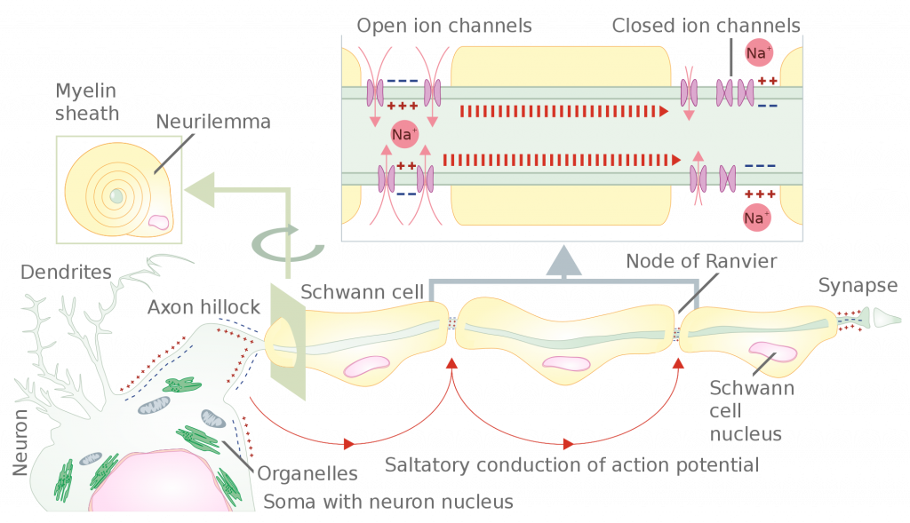
The cell body of the neuron contains the organelles found in a typical animal cell, including the nucleus. It is in the cell body where proteins and neurotransmitter chemicals are made. Dendrites are the branched extensions protruding from the cell body. These carry action potentials to surrounding cells.
The axon is the conductive, long fiber that carries the nervous impulse along the motor neuron.
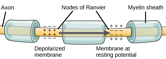
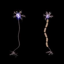
Schwann cells wrap around the axon to form the myelin sheath, which is a lipid and therefore does not allow charged ions or the impulse to pass through it. There are gaps between these myelin sheaths, called nodes of Ranvier.
Resting Potential
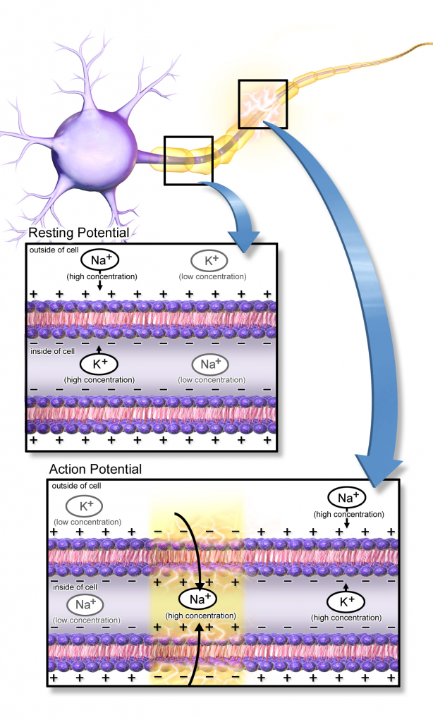
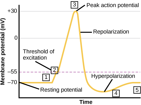
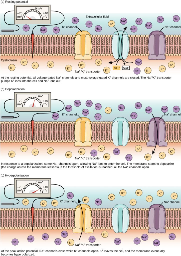
When a neuron is not conducting an impulse, there is a difference between the electrical charge inside and outside of the neuron. This is known as the resting potential.
There are more positive ions, Na+ and K+, outside compared to inside, therefore the inside of the neuron is comparatively more negative at -70mV.
The resting potential is maintained by a sodium-potassium pump, involving active transport and therefore ATP. The pump moves 2K+ ions in and 3 Na+ ions out. This creates a concentration gradient and results in K+ diffusing out and Na+ diffusing in. However, because the membrane is more permeable to K+, more are moved out resulting in the -70mV.
Action Potential
When the potential of the neuron changes, this is an action potential, and it results in a nervous impulse. Changes in membrane permeability lead to this depolarization and generation of an action potential. Throughout depolarization, the Na+ continues to rush inside until the action potential reaches its peak and the sodium gates close. If the depolarization is not high enough to exceed a threshold, then an action potential and the impulse are not produced. This is called the All-or-None Principle. This is important as it ensures that animals only respond to large enough stimuli, rather than responding to every slight change in the environment, which would overwhelm them.
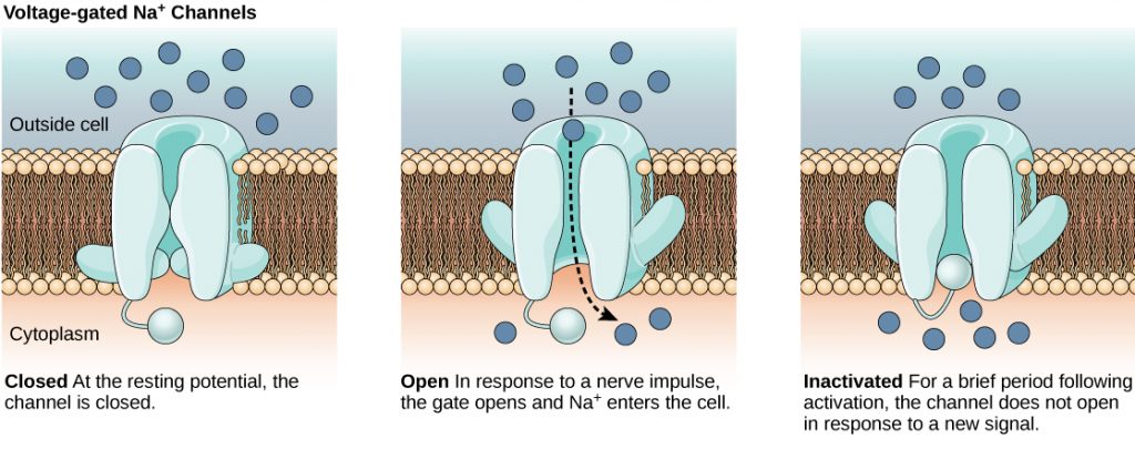
A stimulus provides energy that can cause the sodium voltage-gated channels (proteins in the membrane that only open when a certain voltage is reached) in the axon membrane to open. This causes Na+ to diffuse in, which increases the positivity inside the axon. This causes more voltage-gated channels to open, so even more Na+ diffuses in. When a threshold of +40mV is reached inside the axon, the voltage-gated sodium channels close and instead voltage-gated potassium ion channels open. This results in potassium ions diffusing out, and the axon becoming negative again and repolarizes the axon. Temporarily, the axon becomes more negative than the -70mV and is hyperpolarized. The potassium ion gates will now close, and the sodium-potassium pump restores normal activity to reform the resting potential.
Once an action potential is generated, it moves along the axon like a Mexican wave.
This will occur along the entire axon in a non-myelinated neuron, whereas it is much quicker in myelinated axons. Action potentials can only occur at the nodes of Ranvier, where there is no myelin insulating. Therefore, the localized action potentials will jump from node to node and travel much quicker. This is known as saltatory conduction.
In addition to myelination and saltatory conduction increasing the speed of conduction, temperature and axon diameter also have an impact. The higher the temperature, the faster the ions will diffuse and therefore increase the conduction. The wider the diameter of the axon, the faster the rate of conduction. This is because the larger the diameter, the less likely it is for ions to leak across the membrane and affect the potential.
Refractory Period
Straight after an action potential has been generated, the membrane enters a refractory period when it can't be stimulated because sodium channels are recovering and can't be opened. This means that the voltage-gated sodium channels are closed. This is important for many reasons.
Firstly, it ensures that discrete impulses are produced, meaning that an action potential cannot be generated immediately after another one, and this makes sure that each is separate from another.
Secondly, it ensures that action potentials travel in one direction. This stops the action potential from spreading out in two directions and prevents a response.
Finally, it limits the number of impulse transmissions. This is important to prevent overreaction to a stimulus and therefore overwhelming the senses.
- What is the term given to the potential when the neuron is at rest?
- Your answer should include: resting / potential
- What is the term given to the potential when a stimulus is detected?
- Your answer should include: action / potential
- What is the term given to the period after a stimulus generates a response?
- Your answer should include: refractory / period
- What determines whether the sodium channels within axon membranes open or close?
- voltage
- What is the name of the insulating layer that wraps around an axon?
- Your answer should include: myelin / sheath
- Where in a motor neuron is the nucleus found?
- Your answer should include: cell / body
Synapse & Neuromuscular Junction
Synapses are the gaps between the end of the axon of one neuron and the dendrite of another one. Here the action potential is transmitted as neurotransmitters that diffuse across the synapse. This process occurs as follows: 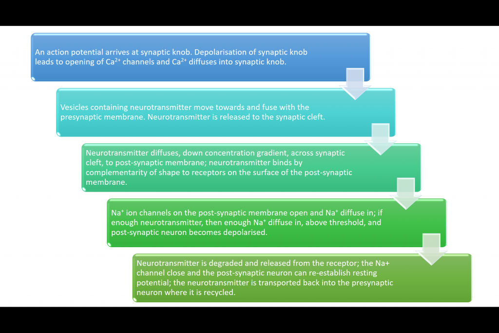 Neuromuscular Junction This is a synapse that occurs between a motor neuron and a muscle and is very similar to a synaptic junction.
| Neuromuscular Junction | Cholinergic Synapse |
| Unidirectional due to the neurotransmitter receptors only being on the postsynaptic membrane | |
| Only excitatory | Could be excitatory or inhibitory. Inhibitory synapses cause chloride ions to move into the postsynaptic neuron and potassium ions to move out. The combined effect of negative ions moving in and positive ions moving out makes the membrane potential increase to -80mV, hyperpolarization, and therefore an action potential is highly unlikely. |
| Connects motor neuron to muscles | Connects two neurons, which could be sensory, relay, or motor. |
| This is the end point for the action potential | A new action potential is generated in the next neuron. |
| Acetylcholine binds to receptors on muscle fiber membranes | Acetylcholine binds to receptors on the postsynaptic membrane of a neuron. |
Summation
Summation is the rapid build-up of neurotransmitters in the synapse to help generate an action potential by two methods: spatial or temporal summation. This is needed because some action potentials do not result in sufficient concentrations of neurotransmitter being released to generate a new action potential.
Spatial summation: many different neurons collectively trigger a new action potential by combining the neurotransmitter they release to exceed the threshold value.
Temporal summation: One neuron releases neurotransmitter repeatedly over a short period of time to add up to enough to exceed the threshold value.
Receptors
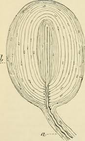
The Pacinian Corpuscle
Receptors detect stimuli, and the Pacinian corpuscle is an example of a receptor.
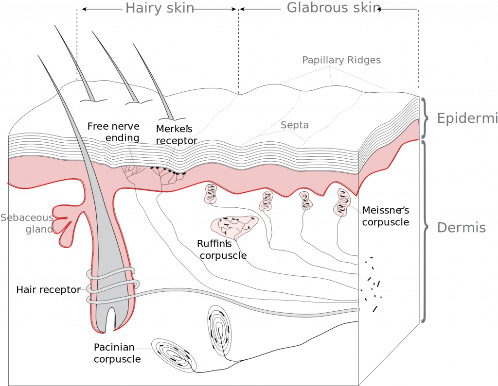
Each receptor responds only to specific stimuli, and this stimulation of a receptor leads to the establishment of a generator potential, which can cause a response. The Pacinian corpuscle responds to pressure changes. These receptors occur deep in the skin, mainly in fingers and feet. It consists of a single sensory neuron wrapped with layers of tissue separated by gel. Plasma membranes contain channel proteins that allow ion transportation. The membranes surrounding the sensory neuron have stretch-mediated sodium channels. In the resting state, Na+ channels are too narrow for Na+ to diffuse, therefore resting potential is maintained. When pressure is applied, it deforms the membrane, stretches and widens the Na+ channels so Na+ diffuses in, which leads to the establishment of a generator potential.
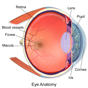
The Human Retina
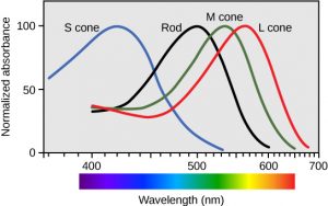
The retina contains two types of photoreceptors: rods and cones.
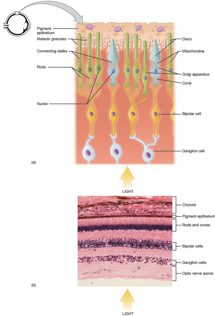
Rods, so called because of their shape, cannot distinguish different wavelengths of light and process images in black and white. Rods can detect light of very low intensity though because many rod cells connect to one sensory neuron. The threshold to create an action potential can be reached even in low light because so many rod cells are connected to a single bipolar cell. This is an example of summation. To create the generator potential, the pigment of rod cells (rhodopsin) must be broken down. There is enough energy from low-intensity light to cause the breakdown. However, this retinal convergence means that the brain cannot distinguish between the separate sources of light that stimulated it. Two dots close together cannot be seen as separate. Rod cells give low visual acuity.
There are three cone cells that contain different types of iodopsin pigment (red, green, and blue), which all absorb different wavelengths of light. Depending on the proportion of each cone cell that is stimulated, we perceive color images. Iodopsin is only broken down if there is a high light intensity, so action potentials can only be generated with enough light.
Usually, only one cone cell connects to a bipolar cell. Therefore, no spatial summation occurs, and cones can only respond to high light intensity, which is why we can't see color when it is dark. As each cone is connected to one bipolar cell, the brain can distinguish between separate sources of light detected. Cone cells give high visual acuity.
The distribution of rods and cones in the retina is uneven. Light is focused by the lens on the part of the retina opposite the pupil, the fovea, which will receive the highest intensity of light. Therefore, most cone cells are located near the fovea, and rod cells are further away.
Control of the Heart
Cardiac muscle is myogenic, but the rate of contraction is controlled by a wave of electrical activity.
The sinoatrial node (SAN) is in the right atrium and is known as the pacemaker. The SAN will release a wave of depolarization across the atria, causing it to contract. The atrioventricular node (AVN) is located near the border of the right and left ventricle within the atria still.
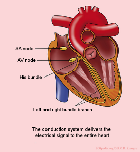

The AVN will release another wave of depolarization when the first reaches it. There is a non-conductive layer between the atria and ventricles, which prevents the wave of depolarization from traveling down to the ventricles. Instead, the bundle of His, running through the septum, can conduct and transmit the wave of depolarization down the septum and the Purkinje fibers in the walls of the ventricles. As a result, the apex and then walls of the ventricles contract. There is a short delay before this happens, while the AVN transmits the second wave of depolarization. This allows enough time for the atria to pump all the blood into the ventricles. Finally, the cells repolarize, and the cardiac muscle relaxes.
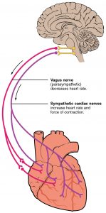
The medulla oblongata in the brain controls the heart rate via the autonomic nervous system. There are two parts: a center linked to the sinoatrial node that increases heart rate via the sympathetic nervous system and another that decreases heart rate via the parasympathetic nervous system.
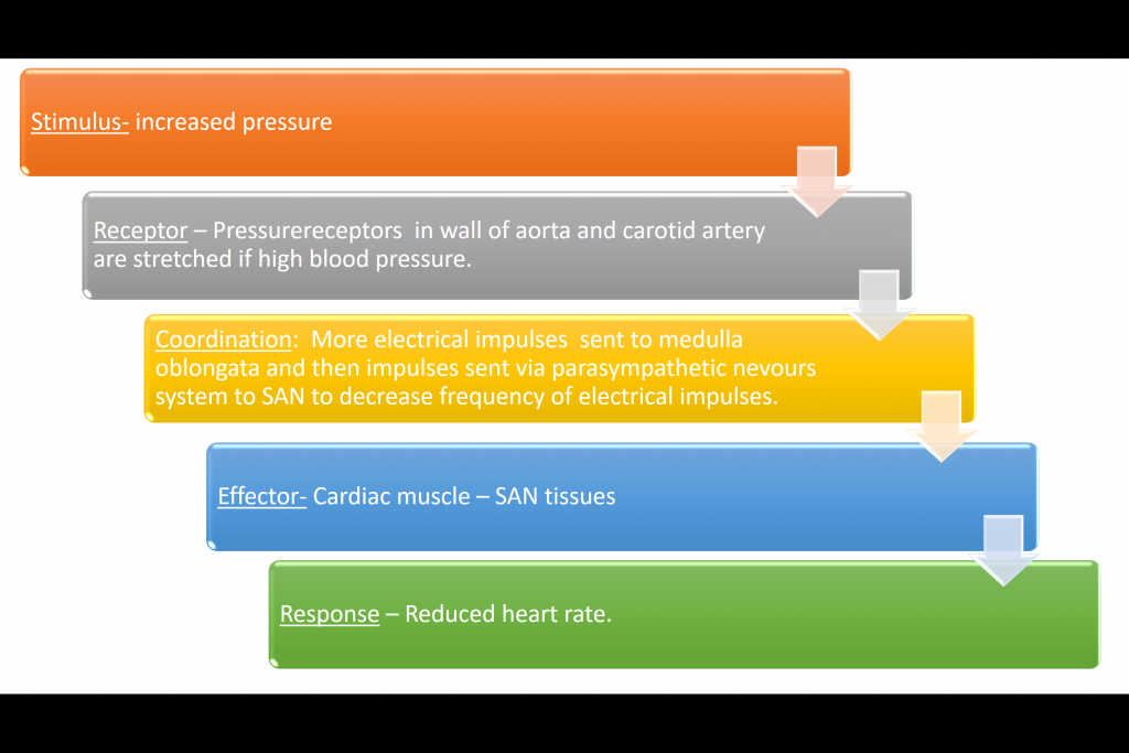
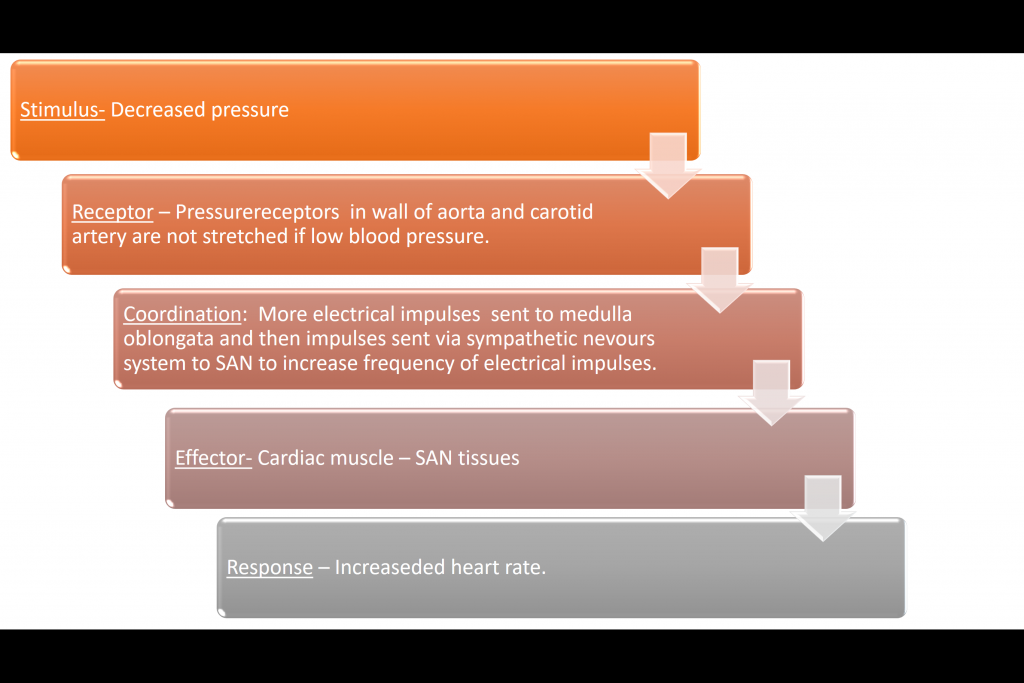
Response to Pressure
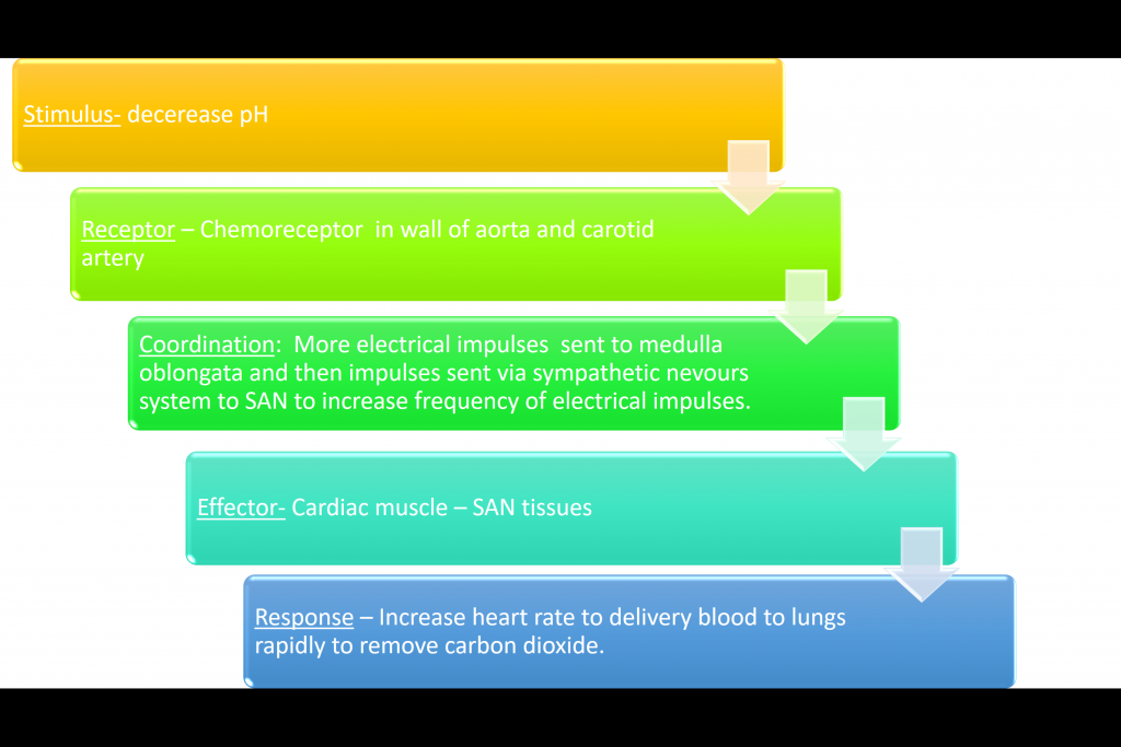
The heart rate changes in response to pH and blood pressure, and these stimuli are detected by chemoreceptors and pressure receptors in the aorta and carotid artery.
Response to pH
The pH of the blood will decrease during times of high respiratory rate due to the production of carbon dioxide or lactic acid.
- What are the two types of summation?
- Your answer should include: spatial / temporal
- What are the names of the two nerves that connect the medulla oblongata and the SAN?
- Your answer should include: sympathetic / parasympathetic
- Where are chemoreceptors located?
- Your answer should include: aorta / carotid / artery
- Where in the retina would you find many cones?
- fovea