Contents
Immunity
Cell Recognition
Cells are labeled with proteins to allow recognition. To prevent your lymphocytes from destroying your own body cells, each of your body cells is labeled with a unique shape protein that your lymphocytes recognize as ‘self’ cells. Any other type of protein detected on the surface of a cell is recognized as ‘non-self’ and is destroyed. This could range from abnormal body cells such as cancer, toxins produced by pathogens, pathogens, and cells from other organisms of the same species.
An antigen is typically a protein molecule on the cell-surface of a membrane of non-self cells. The presence of antigens triggers an immune response and the production of antibodies.
Response to Pathogens
The first line of defense to prevent infection is physical and chemical barriers, such as the skin and stomach acid. If pathogens get past these barriers, then two key types of blood cells respond: the phagocytes and the lymphocytes.
Phagocytosis
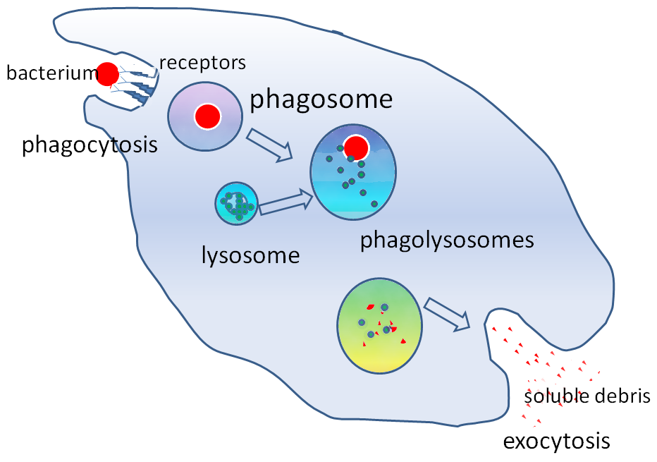
The phagocytes, such as macrophages and neutrophils, travel in the blood and squeeze out of capillaries to engulf and digest pathogens. This process is called phagocytosis and it is non-specific. Damaged cells and pathogens release chemicals that attract the phagocytes to the site of infection.
Phagocytes have receptors that can attach onto chemicals on the surface of pathogens. The phagocyte then engulfs the pathogen into a vesicle to create a phagosome. Within the phagocyte, there are lysosomes that contain hydrolytic lysozyme enzymes.
The lysosome fuses with the phagosome to expose the pathogen to the lysozyme. This hydrolyzes the pathogen, and any soluble useful molecules are absorbed into the cytoplasm of the phagocyte.
The phagocytes will present the antigen of the digested pathogen on their surface; they are then called antigen-presenting cells.
Lymphocytes
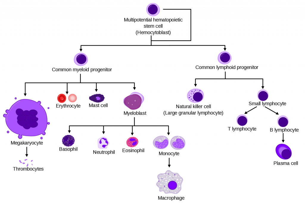
The lymphocytes are part of the specific response to antigens. There are two types: B lymphocytes (B Cells) and T lymphocytes (T cells). Both are created by bone marrow stem cells, but B cells mature in the bone marrow whereas T cells mature in the thymus.
Cell-Mediated Response
This is the response of T cells to a foreign antigen. Receptors on the T cells will bind onto the antigens on antigen-presenting cells and cause the T cell to divide rapidly by mitosis (clonal expansion). These cloned T cells can then differentiate into different specialized T cells:
- Memory T cells to enable a rapid response to reinfection of the same pathogen.
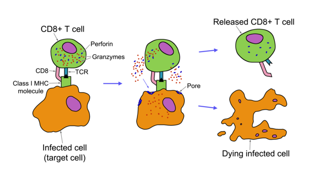
- Cytotoxic T cells kill abnormal cells and infected body cells. They release a protein called perforin which creates pores (holes) in the cell membrane. This allows all substances to move into the cell and causes cell death.
- Helper T cells stimulate B cells to divide and secrete antibodies.
- T cells also stimulate phagocytosis of pathogens.
Humoral Response
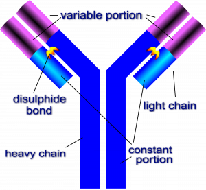
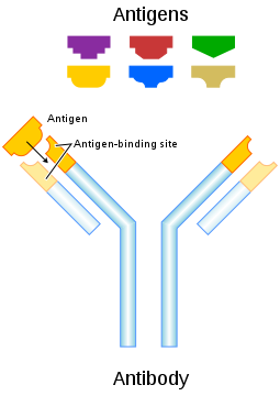
Helper T cells stimulate the B cells and initiate the humoral response, which involves antibodies. Antibodies are proteins that have binding sites complementary in shape to antigens. The binding site is described as the variable region, as the shape changes for each antigen. The rest of the antibody is the constant region. They are made up of four polypeptide chains, two heavy and two light chains. When an antigen binds to an antibody, it is described as an antigen-antibody complex.
Antibodies help destroy pathogens by either causing agglutination or marking. Agglutination is clumping together all the bacterial cells, so it is easier for phagocytes to locate and engulf. They also act as markers to stimulate phagocytes to engulf the cells.